The Age Spot Removal Detail That Changes Everything
Most people treat every brown spot the same — like a cosmetic stain that needs one-size-fits-all removal. That assumption wastes time and can be dangerous. The detail that changes everything is a simple pre-treatment self-screen using an adapted ABCD size rule: it sorts a spot into a harmless solar lentigo, a benign seborrheic keratosis, or a lentigo maligna that demands a biopsy, not a laser.
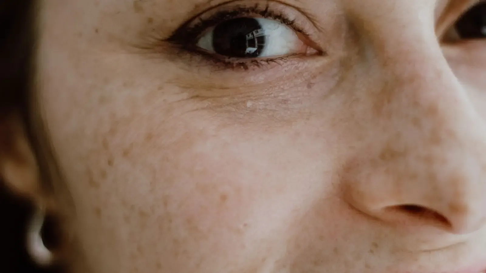
The ABCD Self-Screen: Sorting Brown Spots Before You Treat
Before you book a laser session or buy a spot-fading cream, run every suspicious brown spot through this four-point checklist. The letters stand for Asymmetry, Border, Color, and Diameter. Dermatologists use these same visual markers to decide whether a lesion is safe to treat cosmetically or needs a biopsy. Using them yourself does not replace an in-office skin exam, but it eliminates the cases where a cosmetic procedure would be a clinical mistake.
Asymmetry: One Half Must Mirror the Other
Draw an imaginary line through the middle of the spot. A true solar lentigo — a flat, even-toned sun spot — looks nearly identical on both sides. Seborrheic keratoses stay mostly symmetric despite their irregular surface. Lentigo maligna almost always fails this test: one half differs in shape, color depth, or texture. Asymmetry is the single most decisive sign. A spot that flunks it goes straight to the biopsy-referral tier.
Border: Track It with a Pen Tip
A classic solar lentigo has a crisp border. You can trace its edge without ambiguity. Seborrheic keratoses may have scalloped borders but still appear “stuck on” like wax. A border that fades unevenly into the skin, has notches, or looks smudged points toward lentigo maligna. If the edge resembles a jagged coastline under magnification, that spot needs a dermatologist’s dermoscope, not a laser pulse.
Color: Count the Shades Inside the Spot
Solar lentigines hold one color: light tan, medium brown, occasionally dark brown — uniform. Seborrheic keratoses can range from flesh-tone to near-black, but typically read as one dominant hue with a waxy finish. Lentigo maligna mixes colors: tan, brown, black, sometimes pink or gray in the same lesion. That pigment variegation is a red flag. Two or more distinct colors in one spot override any cosmetic plan.
Diameter: Size Has a Threshold
Use a millimeter ruler. Solar lentigines are usually under 6 mm — the size of a pencil eraser. Seborrheic keratoses can grow larger and still be benign, but their raised, warty nature distinguishes them. Lentigo maligna often starts small and grows slowly; any flat brown patch larger than 6 mm that also shows asymmetry, irregular borders, or color variegation must be evaluated. Size alone is not the verdict; it becomes meaningful only with the other ABCDs.
Quick Reference: Solar Lentigo vs. Seborrheic Keratosis vs. Lentigo Maligna
| Feature | Solar Lentigo | Seborrheic Keratosis | Lentigo Maligna |
|---|---|---|---|
| Symmetry | Symmetric | Mostly symmetric | Asymmetric |
| Border | Sharp, well-defined | Often scalloped, “stuck-on” | Irregular, smudged, notched |
| Color | Uniform light to medium brown | Uniform, may be dark or waxy | Variegated: mixed browns, black, pink |
| Diameter | Usually <6 mm | Can be >6 mm, warty growth | Often >6 mm, enlarging over time |
| Surface | Flat | Raised, warty, greasy feel | Flat, sometimes scaly |
| Next Step | Cosmetic removal safe | Cosmetic removal possible; confirm benign | Biopsy referral |
The table is a sorting tool, not a diagnosis. If your spot lands in the right-hand column, pause every cosmetic plan and schedule a medical skin check.
Matching the Spot to the Corrective Tier
Once the self-screen places a brown spot solidly in the solar lentigo or benign seborrheic keratosis category, treatment follows a three-tier logic that board-certified dermatologists apply daily.
Tier 1: Laser for Flat, Uniform Solar Lentigines
Pigment-specific lasers — Q-switched Nd:YAG, alexandrite, or picosecond devices — shatter melanin in the epidermis with minimal thermal damage. For Fitzpatrick skin types I-III, they clear a spot in one or two sessions. In skin of color (types IV-VI), the risk of post-inflammatory hyperpigmentation (PIH) forces a different protocol: lower fluence, 1064 nm wavelength that spares epidermal melanin, and mandatory test spots. A provider who skips the test spot on darker skin is taking an unacceptable risk. No laser should touch a spot that hasn’t passed the ABCD screen; blasting an unrecognized lentigo maligna can mask progression.
Tier 2: Chemical Peels for Solar Lentigines and Thin Seborrheic Keratoses
A medium-depth peel — 35% TCA or a Jessner’s–TCA combination — lifts sun-damaged pigment by removing superficial epidermal layers. Peels carry a wider safety margin for darker skin when used at lower concentrations, but pre-treatment with a topical retinoid or pigment suppressor (hydroquinone 4% or azelaic acid) is not optional; it cuts PIH risk sharply. For thin seborrheic keratoses, TCA can crust them off; thick, deeply pigmented SKs often need cryotherapy or shave excision performed by a dermatologist, not a peelist. Choose tier 2 when laser is contraindicated or cost is the barrier.
Tier 3: Biopsy — When the Spot Raises Any Doubt
If the ABCD screen flags a problem, or a provider sees something suspicious under dermoscopy, the lesion jumps to tier 3. A biopsy — punch, shave, or excisional — is the only definitive path. No laser, no peel, no home kit can rule out lentigo maligna or melanoma. The decision is binary: if there’s even a low probability of malignancy, the correct treatment is a biopsy, period.
The Fitzpatrick Factor: Skin Tone Governs Safety, Not Just Efficacy
The same solar lentigo on a Fitzpatrick type II and a type V patient requires two completely different plans. Darker skin’s melanocytes react aggressively to injury; a laser pulse or peel can trigger a dark post-inflammatory mark worse than the original spot. So tier selection depends as much on the skin it sits on as on the lesion itself.
For solar lentigines in skin of color, the safer route is often a series of superficial glycolic acid peels or low-energy, fractionated 1064 nm Nd:YAG treatments. Pre-treat with a tyrosinase inhibitor for weeks — no exceptions. A test spot behind the ear before facial treatment is mandatory. For seborrheic keratosis removal in darker skin, cryotherapy or shave excision can work, but strict sun protection and melanin-suppressing topicals afterward are required. If a med spa’s “age spot laser package” doesn’t factor in your Fitzpatrick type, walk out. The ABCD screen sorts the lesion; the Fitzpatrick scale selects the safe device and protocol.
When a Spot Is More Than a Spot: Red Flags That Cancel Cosmetic Plans
A brown spot that changes over weeks, itches, bleeds, or develops a nodule — it is not an aging issue. Lentigo maligna can mimic a solar lentigo for years before becoming invasive melanoma. The ABCD screen catches the static red flags: asymmetry, color variegation, diameter >6 mm with irregular borders. But a changing or evolving spot is the strongest alarm. Dermatologists also look for the “ugly duckling” sign — the one that doesn’t match the rest.
Spot any of these, and the only action is a dermatology appointment. No IPL, no cryopen, no at-home freezing kit. A board-certified dermatologist might use dermoscopy to examine pigment networks; if suspicion remains, a biopsy is done under local anesthesia. The pathology report dictates everything else.
Key Takeaways
- The adapted “ABCD size” rule (Asymmetry, Border irregularity, Color variegation, Diameter >6 mm) categorizes brown spots into cosmetic solar lentigines, benign seborrheic keratoses, and lentigo malignas requiring biopsy.
- A spot that passes the ABCD screen — symmetric, sharp border, uniform color, under 6 mm — qualifies for tier 1 (laser) or tier 2 (chemical peel) removal.
- Seborrheic keratoses often are symmetric but large; they’re benign and removable via peel, cryotherapy, or shave excision after confirmation.
- Fitzpatrick skin types IV-VI demand a modified protocol: lower-energy lasers, pre-treatment lightening agents, and test spots to prevent PIH.
- Any lesion failing the ABCD screen — or any changing, symptomatic spot — bypasses cosmetics entirely and goes to tier 3: biopsy referral.
The Next Step After a Clean ABCD Self-Screen
If your spot matches the solar lentigo column and your skin type is compatible with the planned treatment, book a consultation with a board-certified dermatologist — not a walk-in laser lounge. The provider will confirm your self-screen with dermoscopy, exclude occult lentigo maligna, and set the laser or peel parameters to your skin. If even one ABCD feature leans toward the lentigo maligna side, schedule a medical dermatology appointment immediately. The detail that changes everything — that quick ABCD scan — is not a substitute for a professional exam. It’s the filter ensuring your quest for even-toned skin doesn’t accidentally ignore a lesion that belongs under a pathologist’s microscope, not a laser’s beam.
Frequently Asked Questions
What is the ABCD rule for age spots?
An adaptation of melanoma screening: check Asymmetry, Border irregularity, Color variation, and Diameter larger than 6 mm. Solar lentigines are symmetric with sharp borders and uniform color; lentigo maligna breaks multiple rules.
Can seborrheic keratosis be removed at home?
No. Home acids, freezing kits, or picking risk infection, scarring, and mask a possible malignancy. Dermatologists remove SKs safely with cryotherapy, curettage, or shave excision.
Is laser safe for all skin tones?
Not equally. Darker skin (Fitzpatrick IV-VI) risks post-laser hyperpigmentation. Safe treatment requires conservative settings, longer wavelengths, a test spot, and pre-treatment lightening. A provider skilled in skin of color is essential.
How do I know if my brown spot is cancerous?
Apply the ABCD rule: asymmetry, irregular borders, color variegation, diameter >6 mm, plus any change or symptom. If present, get a dermatologist’s dermoscopy and possible biopsy.
Can I use the ABCD rule on spots I’ve had for years?
Yes. Long-standing spots can still turn malignant, especially if they evolve. Run the ABCD checklist; a stable spot that passes is likely benign, but any new change demands an exam.
What does a lentigo maligna look like?
A flat, enlarging brown patch with multiple shades of tan, brown, black, irregular borders, and asymmetry. Common on sun-exposed areas in older adults.
Pigment Curious
We are people who want to better understand skin pigmentation, dark spots, and how different skin tones respond to skincare and professional treatments. We look beyond simple beauty claims, focusing on scientific evidence, clear explanations, and practical knowledge that helps people make more informed decisions about their skin.
This is the clearest explanation of the ABCD rule I’ve ever read. I’ve had a flat brown spot on my cheek for years and always assumed it was just an age spot, but now I’m going to check the symmetry and border tonight. Thank you for making it so approachable!
Great article. I’m a Fitzpatrick type V and had a bad experience with a laser test spot that left a dark mark that took months to fade. Are there any specific at-home pre-treatments you’d recommend before trying a low-energy peel for a solar lentigo on my forehead?
Thank you for sharing that, John — and I’m sorry you had that experience. For Fitzpatrick V, pre-treating with a tyrosinase inhibitor like 4% hydroquinone or azelaic acid for at least 4–6 weeks is key. Always have a board-certified dermatologist supervise the peel, and insist on a test spot behind the ear first. It makes all the difference.
I used the ABCD check on a spot behind my ear that I’d ignored for ages. It was slightly asymmetric and had two shades — turned out to be a lentigo maligna after biopsy. Caught early, no invasion. This article should be in every dermatology waiting room.