Alexandrite Laser for Spots: The Skin Type That Must Avoid It
If you are searching for an Alexandrite laser to clear pigmented spots, the data leaves no room for doubt: Fitzpatrick skin types IV, V, and VI must not be treated with this device. The 755-nm wavelength that removes discrete spots on light skin becomes non-selective thermal damage on melanin-rich skin—burns, permanent hyperpigmentation, and textural scarring replace any cosmetic benefit. This is not a matter of adjusting fluence or spot size; it is a binary exclusion.
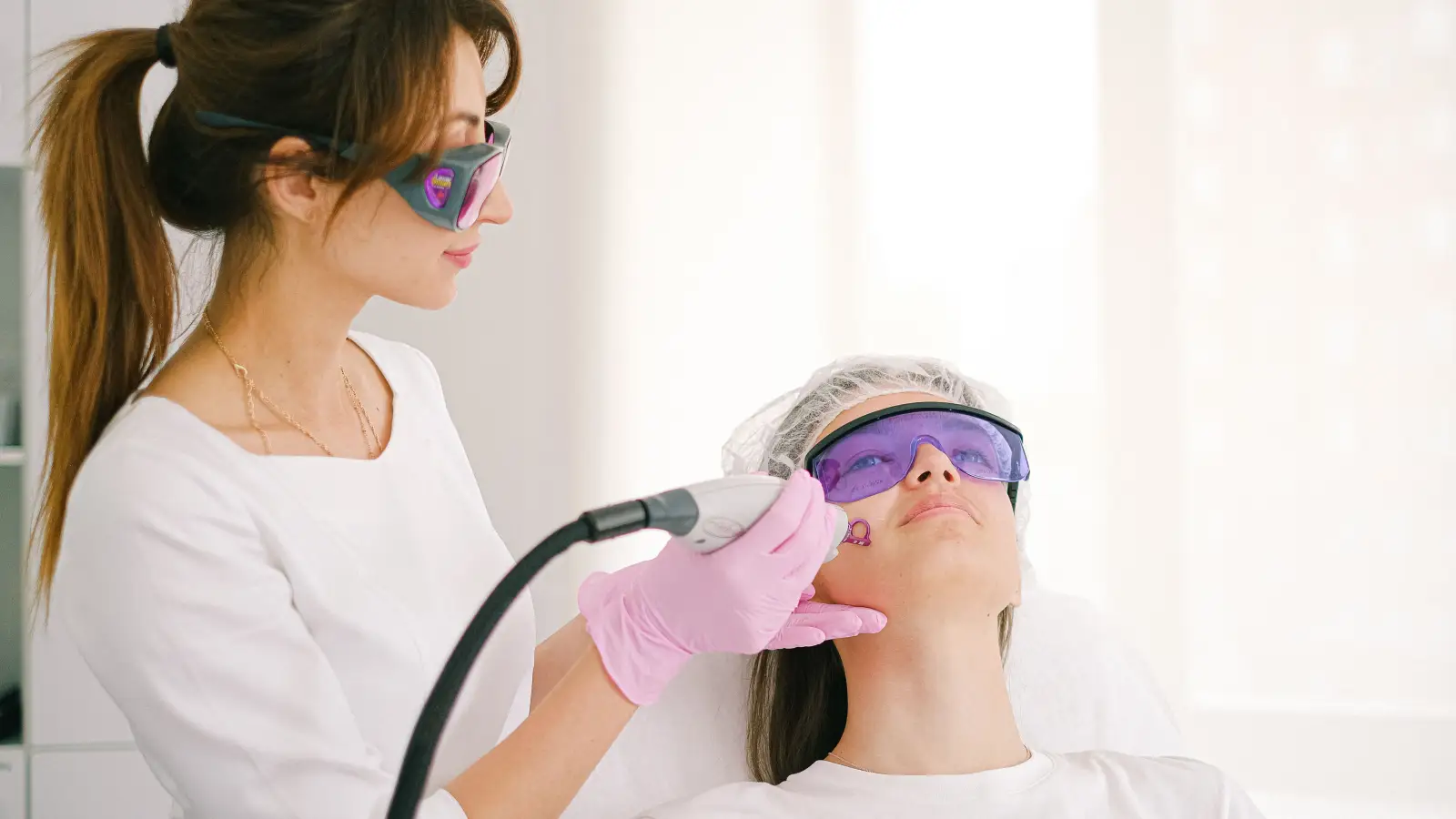
The pre-appointment screening checklist that follows is built on that hard stop and layered with every additional contraindication a dermatologist would flag: recent ultraviolet exposure, melasma, a personal history of keloid scar formation, and a short list of medication and physiological states that render the laser risk unacceptable. Read it as if you were evaluating a patient, because in this context, you are.
Fitzpatrick IV–VI Is an Absolute Contraindication
The Fitzpatrick phototype scale categorizes skin by its response to ultraviolet radiation. Types I and II burn and do not tan; type III may burn then tan; type IV tans readily and rarely burns; type V tans deeply; type VI is deeply pigmented. Laser dermatology uses this scale as a proxy for epidermal melanin density. At 755 nm, Alexandrite light is absorbed powerfully by melanin. In a fitzpatrick I–III spot, that melanin is concentrated inside the lesion. In types IV and above, the surrounding normal skin contains enough competitive melanin to pull the thermal injury outside the target border.
The result is not spot removal. It is widespread thermal destruction of melanocytes, often with paradoxical hyperpigmentation or depigmentation. No conservative parameter adjustment removes this risk. The physics of the wavelength itself makes darker skin an absolute contraindication. If a provider offers Alexandrite to a skin type V patient on the grounds of a “test patch,” that provider is operating outside the standard of care.
Recent Sun Exposure Resets the Risk Profile
Ultraviolet exposure activates melanocytes and increases melanin production in the epidermis for days to weeks after any visible tan fades. That activation temporarily shifts the skin’s behaviour toward a higher Fitzpatrick category. A patient who is ordinarily a safe type III can sustain the same thermal burn a type V would, if they present with a recent weekend tan or sub-acute sunburn.
The standard pre-procedure window is a full four to six weeks of rigorous photoprotection. Even then, a Wood’s lamp examination can reveal latent pigment that gross examination misses. Any detectable tan in the treatment field should postpone the session. The risk of post-inflammatory hyperpigmentation from a laser on a recently tanned skin is fully preventable—by waiting.
Melasma Is Not a Simple Pigmented Lesion
Melasma looks like hyperpigmentation but behaves like a hormonally mediated, heat-sensitive condition. It flares with inflammation and thermal energy. The 755-nm wavelength of the Alexandrite laser penetrates to the dermal-epidermal junction, precisely where melasma resides, and deposits the exact type of heat that triggers a darkening cascade. What was a faint patch can become a deep, recalcitrant brown.
This contraindication holds for all Fitzpatrick types. The diagnostic step before any laser consultation for “spots” must be to rule out melasma. If melasma is present, the treatment pathway diverges entirely: rigorous sunscreen with visible light protection, topical tranexamic acid or hydroquinone, and possibly fractional 1927-nm thulium non-ablative resurfacing by an experienced dermatologist—never Alexandrite.
Keloid and Hypertrophic Scarring History Are a No
A laser pulse is a controlled thermal wound. In a patient with a history of keloid or hypertrophic scar formation, that controlled wound heals by overproducing collagen, yielding a raised, itchy, often larger scar. This can occur regardless of skin color. Even on a Fitzpatrick II background, a small lentigo removal can transform into a keloid if the patient’s healing biology is primed for it.
The screening question is straightforward: Do you tend to form raised scars after cuts, piercings, or surgical incisions? If yes, Alexandrite spot removal is not worth the risk. Alternative modalities—cryotherapy, topical fading agents, or excision with close monitoring—must be weighed instead.
Additional Screening Exclusions
The full checklist includes other disqualifiers that are less widely discussed but equally consequential:
Photosensitizing Medications and Supplements
Tetracyclines, fluoroquinolones, St. John’s Wort, and certain retinoids increase the skin’s absorption of light and lower the threshold for thermal injury. A patient taking any of these is not a candidate until a washout period—often two to four weeks—has been completed.
Unstable Pigment Disorders
Active vitiligo, lichen planus pigmentosus, or fresh post-inflammatory hyperpigmentation from a prior procedure create an unpredictable pigment background. The laser can trigger new areas of depigmentation or worsen the existing dyschromia. No procedure should be performed on a moving target.
Pregnancy and Lactation
Hormonal shifts during pregnancy alter melanocyte activity, making skin less predictable. While no direct evidence of fetal harm exists for cutaneous lasers, the consensus is to defer all non-essential procedures until after breastfeeding. This is a precautionary exclusion, not a pharmacological one.
Gap Between Expectation and Reality
A spot that can be erased cleanly is one thing. A spot that a patient has scrutinised to the point of body dysmorphic disorder is another. When the psychological weight attached to a lesion outweighs what any laser can deliver, a responsible dermatologist will decline to treat, regardless of how technically “safe” the skin type appears.
The Self-Screening Checklist
Before contacting a clinic, answer these questions. A single “yes” means Alexandrite should be off the table.
- Is your natural, untanned skin Fitzpatrick type IV, V, or VI?
- Has the treatment area had a sunburn or visible tan within the last six weeks?
- Is there any history of melasma on the treatment area?
- Do you form raised keloid or hypertrophic scars after minor injuries?
- Are you currently taking any antibiotic (tetracycline, doxycycline, ciprofloxacin), oral retinoid, or St. John’s Wort?
- Have you had another laser, peel, or microneedling on the same area that still shows redness or pigmentation change?
- Are you pregnant or breastfeeding?
If you checked a box, the next step is not to find a provider willing to overlook it. It is to schedule a consultation with a board-certified dermatologist who can evaluate the lesion itself and discuss the correct alternative for your skin—likely a 1064-nm Nd:YAG or a topical regimen, depending on the diagnosis.
Key Takeaways
- Alexandrite laser is absolutely contraindicated for Fitzpatrick IV–VI; no fluence adjustment changes the physics of 755-nm absorption in melanin-dense skin.
- Even a mild recent tan temporarily elevates risk to levels seen in darker skin types; a four-to-six-week sun-avoidance window is mandatory.
- Melasma should never be treated with Alexandrite; it must be ruled out as a diagnosis before any laser is considered.
- Keloid formers should avoid any procedure that creates a thermal wound in the dermis.
- Photosensitizing drugs, unstable pigment conditions, and pregnancy are additional hard stops.
- A structured self-screening checklist can prevent a dangerous consultation from ever taking place.
The checklist is not a substitute for dermoscopy and a trained eye. Some lesions—seborrhoeic keratoses, atypical nevi, lentigo maligna—can masquerade as simple spots. A board-certified dermatologist should examine every lesion before it is treated. The safest use of this information is to walk into that appointment already knowing which laser is not for you.
Pigment Curious
We are people who want to better understand skin pigmentation, dark spots, and how different skin tones respond to skincare and professional treatments. We look beyond simple beauty claims, focusing on scientific evidence, clear explanations, and practical knowledge that helps people make more informed decisions about their skin.