How Dermatologists Match PIH Treatment to Spot Depth & Skin Tone
Dermatologists do not treat PIH by trial and error. They match treatment to the exact depth of the pigment and the patient’s Fitzpatrick skin type, using a decision tree that always starts with topical therapy and only escalates when the risk of causing more pigmentation is controlled. After reading, you will understand that systematic logic—and know which questions to bring to a consultation.
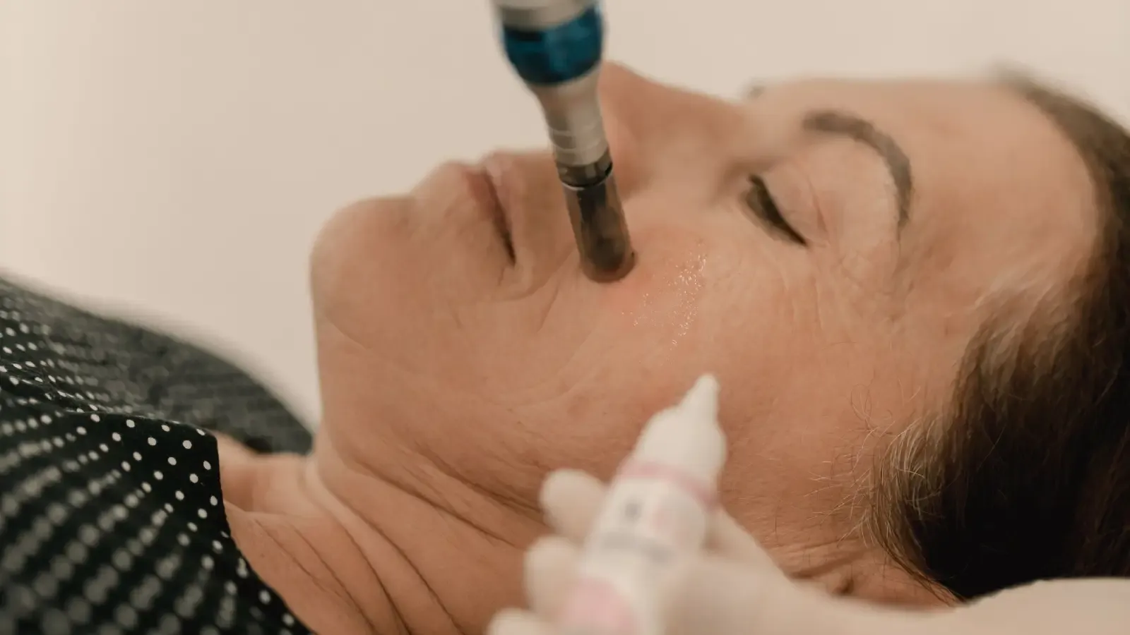
The algorithm is straightforward: identify whether the discoloration is epidermal, dermal, or mixed; then select the least aggressive intervention capable of reaching that depth without provoking post-inflammatory hyperpigmentation in a given skin tone. Every treatment decision beyond that follows a strict hierarchy.
The First Branch: Spot Depth Determines What Can Work
Epidermal PIH: Where Topicals Reach the Target
Epidermal pigment sits in the uppermost living layer of skin. It looks brown, well-defined, and darkens under a Wood’s lamp. Acne, minor wounds, and superficial inflammation produce this pattern. Because the pigment lies within reach of permeating agents, topical therapy is effective. Tyrosinase inhibitors—hydroquinone, azelaic acid, kojic acid—suppress new melanin. Retinoids accelerate keratinocyte turnover, sloughing pigment-bearing cells. Exfoliants physically remove the stained layer.
No procedure is needed here as a first response. When topicals work, the decision tree stops expanding. That alone spares a significant number of patients from unnecessary risk.
Dermal PIH: Depth That Defies Most Topicals
Dermal PIH results from deep inflammation that pushed melanin into macrophages, where it lingers. The clinical appearance is blue-gray, slate, or muddy, with no enhancement under Wood’s light—sometimes it becomes less obvious. This geography resists almost all topical agents: they cannot descend to the dermal-epidermal junction in sufficient concentration to dismantle trapped pigment. Hydroquinone and retinoids may lighten the epidermal component but leave the dermal core untouched. Tranexamic acid, topical or oral, might quiet the inflammatory signals that perpetuate the spot, but clearance is partial and slow.
For purely dermal PIH, dermatologists accept that months of topical treatment often serve only to rule out non-invasive routes before considering devices. The algorithm uses that waiting period deliberately—to confirm that no safer option will suffice.
Mixed PIH: When Both Layers Are Involved
Most problematic spots are mixed. The brown hue sits on top; a blue-gray undertone signals dermal involvement. Treating mixed PIH means tackling the epidermal portion fully first, then evaluating what remains. Topical combinations can often clear the superficial layer. The residual dermal component then determines if the patient moves deeper into the decision tree. This sequential approach prevents over-treatment of the epidermis when the real problem lies below it.
Fitzpatrick Skin Type: Calculating the Risk of Post-Treatment PIH
Melanocyte biology changes across Fitzpatrick types. Types I and II have low melanocyte reactivity; aggressive treatments rarely trigger rebound pigmentation. Types III and IV present moderate risk; types V and VI carry high risk because the melanocytes are already primed for overproduction. This gradient is not a suggestion—it is the single most important risk factor when deciding whether to peel or laser.
A Fitzpatrick VI patient with epidermal PIH will not receive a 50% glycolic acid peel, even if it could clear the spots quickly. The probability of post-peel PIH is too high. The algorithm responds by maximizing topical therapy: compounded hydroquinone, retinoid, possibly a low-potency corticosteroid, for months. If that fails, a 20% mandelic acid peel with a test spot might be considered. For dermal PIH in the same skin type, lasers are approached with extreme restraint—low fluence, long wavelength, and multiple test spots observed for 8 weeks—and only when the quality-of-life impact justifies the gamble.
A Fitzpatrick I patient with dermal PIH, by contrast, may advance to a fractional non-ablative laser sooner because the safety buffer is wider. The decision tree adjusts all escalation thresholds according to this melanin-determined risk.
Building the Topical Foundation: First-Line Agents Matched to Depth
The Universal Opening
No branch begins with a peel or a laser. A structured topical regimen is always the first step, serving two functions: it treats the most accessible pigment and it reveals how the skin tolerates active ingredients before any trauma is introduced.
For epidermal PIH across all skin types, the standard foundation includes a tyrosinase inhibitor (hydroquinone 4%, azelaic acid 15–20%, or cysteamine), a retinoid (tretinoin 0.025–0.05%, adapalene 0.1–0.3%), and a broad-spectrum tinted mineral SPF 30+ every morning. Visible light protection is especially relevant for darker skin tones.
When dermal involvement is suspected, dermatologists layer in topical tranexamic acid 3–5% or niacinamide 4–5% in the morning routine, and may add oral tranexamic acid for widespread, stubborn spots. The logic: these agents don’t dissolve dermal pigment, but they lower the inflammatory persistence that keeps it in place, giving the body’s clearance mechanisms a chance.
Calibrating to Skin Tone Risk
Fitzpatrick III–VI patients cannot tolerate high-concentration leave-on acids as daily exfoliants without risking irritation-induced PIH. The topical base substitutes glycolic acid with mandelic acid, lactic acid, or polyhydroxy acids. Azelaic acid is favored not only for its pigment inhibition but for its anti-inflammatory effect. The aim is sustained pigment suppression with near-zero irritation.
Chemical Peels in the Algorithm: When Topicals Plateau and How to Match Peel Type to Depth and Skin Tone
The Decision Threshold
A peel enters the algorithm only after 12–16 weeks of consistent topical use with an insufficient response—typically for epidermal or mixed PIH. The peel’s agent, strength, and application are chosen to add just enough depth of injury to reach the pigment without crossing into the reticular dermis, where healing becomes unpredictable.
Fitzpatrick I–II, epidermal PIH: 30–50% glycolic acid or 20–30% salicylic acid peels usually work. Fitzpatrick III–IV: the safer options are 20–30% mandelic acid or 20% salicylic acid, or very light glycolic starting at 20%; mandelic’s larger molecular size slows penetration and reduces irritation. Fitzpatrick V–VI: even a 20% mandelic acid peel is often preceded by a test spot and a long observation period. Some clinicians avoid peels altogether in this group unless topical failure is severe and the patient is fully informed.
Dermal PIH is not a peel target. Peels reach the epidermis and, at best, the papillary dermis. The deeper the peel needed to address dermal pigment, the greater the risk of scarring and permanent dyspigmentation in skin of color. The decision tree routes dermal PIH past peels entirely, toward the device branch.
Non-Negotiable Safety Steps
- A test spot on a hidden area, observed for 4 weeks, is mandatory for any peel in Fitzpatrick III and above.
- Pre-peel priming with hydroquinone for 2–4 weeks reduces the melanocyte response that causes post-peel PIH.
- Strict sun protection with tinted mineral SPF during recovery is required; even incidental UV exposure can reignite pigmentation.
Laser and Energy-Based Devices: The Final Branch for Stubborn Dermal PIH
The Gating Criteria
Energy devices are reserved for PIH that has not responded to 6–12 months of topical therapy and, where applicable, superficial peels—and only when the remaining pigment is confirmed dermal or mixed with a dominant dermal component. The gate is both depth and a cold-eyed risk-benefit calculation.
Device Selection by Depth and Skin Tone
The safest wavelength for dermal PIH in melanin-rich skin is 1064 nm, which transmits through the epidermis with minimal melanin absorption, then fragments the deeper pigment via photoacoustic effect. Low-fluence Q-switched Nd:YAG and picosecond 1064 nm lasers are the go-to tools. The pico variant’s ultra-short pulses create mostly mechanical disruption rather than heat, lowering thermal injury.
For Fitzpatrick I–II, low-fluence Q-switched ruby (694 nm) or alexandrite (755 nm) lasers may be used, but these shorter wavelengths carry higher melanin affinity and are avoided in darker skin. Fractional non-ablative lasers (1550 nm, 1927 nm) can be deployed in Fitzpatrick I–IV for mixed PIH, but conservative energy settings are critical.
Intense pulsed light is almost never the right choice for PIH in skin of color. Its broad spectrum heats epidermal melanin unpredictably, frequently causing paradoxical darkening. The decision tree selects a laser over IPL every time.
Protocol for the Highest-Risk Skin Types
For Fitzpatrick V–VI, a dermatologist follows a strict sequence:
- Perform a small test spot with the lowest fluence that yields clinical effect and observe for 6–8 weeks.
- Use cooling, avoid overlapping pulses, and limit the test area.
- Pair the laser with a tyrosinase inhibitor before and after treatment to suppress melanogenesis.
- Space sessions at least 4–6 weeks apart and stop if no progress, rather than pushing through a fixed number.
From Assessment to Action: The Dermatologist’s Sequential Pathway
Step-by-Step Decision Logic
- Determine PIH depth. Wood’s lamp and history. Epidermal PIH enhances; dermal PIH does not or fades under the light. Mixed shows partial enhancement.
- Classify Fitzpatrick skin type. The phototype sets the risk ceiling for any interventional step.
- Start topical therapy immediately, matched to depth.
– Epidermal: tyrosinase inhibitor + retinoid at night; azelaic acid or niacinamide in the morning; tinted mineral sunscreen.
– Dermal: add topical or oral tranexamic acid; consider low-concentration retinoid for penetration without irritation.
- Reassess at 12–16 weeks.
– >50% improvement in epidermal marks: continue topicals. Slower progress: add a gentle chemical exfoliant.
– Dermal marks with no change: remain on optimized topicals. Only after 6 months of zero progress consider moving forward.
- Epidermal PIH, still unsatisfactory: introduce a superficial peel on the basis of Fitzpatrick risk. Mandelic acid for III–VI, glycolic acid for I–II. Prime with hydroquinone and test first.
- Dermal PIH, persistent: perform a low-fluence 1064 nm Q-switched or pico laser test spot on a hidden area; observe 8 weeks. Proceed only if no PIH arises. Tailor fluence, spot size, and intervals to the individual’s sensitivity.
- Maintenance indefinitely: retinoid or azelaic acid plus daily sunscreen, because any minor inflammation can re-initiate PIH in susceptible skin.
PIH Treatment Matching at a Glance: Depth, Skin Tone, and Escalation Table
| PIH Depth | Fitzpatrick I–II | Fitzpatrick III–IV | Fitzpatrick V–VI |
|---|---|---|---|
| Epidermal | Topicals (HQ, retinoid, AHA serum). If slow, 30–50% glycolic peel. | Topicals (azelaic acid, retinoid, mandelic or lactic exfoliant). If needed, 20% mandelic or salicylic acid peel. | Topicals only for 4–6 months. Test spot 20% mandelic peel if essential. Avoid glycolic/TCA. |
| Mixed | Topicals + superficial glycolic or salicylic peels after 12 weeks. | Topicals + cautious mandelic peels after 16 weeks. Add oral TXA if dermal component visible. | Topicals (add oral TXA early). Avoid peels if possible. Consider only mandelic with test spot and long observation. |
| Dermal | Topicals (HQ + retinoid + oral TXA). If unchanged, low-fluence QS 1064 nm or pico laser after 6 months. | Topicals + oral TXA. Fractional non-ablative laser or low-fluence 1064 nm with extreme caution; test spot mandatory. | Prolonged topicals + oral TXA. Low-fluence 1064 nm pico laser only by expert, with multiple test spots and 8-week observation; consider deferring if risk outweighs benefit. |
Common Questions About PIH Depth and Treatment Matching
How can I tell if my PIH is epidermal or dermal?
A Wood’s lamp exam by a dermatologist is the objective standard. Epidermal PIH darkens under the light; dermal PIH does not. At home, brown color indicates epidermal, blue-gray hints at dermal. Mixed spots show both. Accurate depth determination is non-negotiable because it forecasts which treatments have any chance of working.
Why do dark skin tones need a different approach for PIH treatment?
Melanocytes in darker skin are more numerous, larger, and highly responsive to any trigger. Even minimal procedural trauma can ignite a cascade of excess melanin production, leaving the patient with more pigment than before. The decision tree protects against this by demanding maximum topical effort first, lower-strength peels, and laser protocols that prioritize epidermal bypass and cooling.
Can hydroquinone treat dermal PIH?
Hydroquinone inhibits tyrosinase in the epidermis. It does not break down melanin already trapped in dermal macrophages. It can suppress ongoing pigment transfer and help with an epidermal overlay, but it will not resolve truly dermal spots. That limitation is why dermal PIH so often leads to the device branch of the algorithm.
Are there any lasers safe for Fitzpatrick VI skin with PIH?
Only low-fluence 1064 nm Q-switched or picosecond lasers, used by providers with deep experience in skin of color. Even then, safety is earned through multiple test spots observed for 6–8 weeks—no rush, no shortcuts. The decision is never automatic; it rests on a judgment that the untreated spot is worse than the risk of a procedure.
How long does it take to see results from topical treatment for PIH?
Epidermal PIH may lighten in 8–12 weeks, with substantial clearance at 16–24 weeks. Dermal PIH topicals often require 6 months to show any change, and complete clearance is rare. Consistency and daily sunscreen use are the only ways to accelerate the timeline; skipping either resets the clock.
Matching Treatment to Depth and Skin Tone: Core Principles
- PIH depth sorts the entire treatment path. Epidermal pigment responds to many agents; dermal pigment resists all but the most deliberate interventions.
- Fitzpatrick skin type is not a label—it is a predictive variable that caps how aggressively you can treat without causing harm.
- Topicals are always the first node because they buy time, build tolerance, and expose which pigment component is truly refractory.
- Chemical peels enter only when topicals plateau, and for darker skin, the peel choice shifts entirely to mandelic acid or similar low-irritation agents, if used at all.
- Lasers are the terminal node, never the starting point, and in high-risk skin types they are deployed with a level of caution that makes clear refusal an equally valid option.
The single most important step before treating PIH is determining whether the pigment is epidermal, dermal, or mixed—because that answer determines what topicals can do and when the risk of procedures may be justified. Knowing this, and understanding precisely how your Fitzpatrick skin type amplifies or dampens risk, allows you to approach any treatment decision with a clear head rather than hope.
Pigment Curious
We are people who want to better understand skin pigmentation, dark spots, and how different skin tones respond to skincare and professional treatments. We look beyond simple beauty claims, focusing on scientific evidence, clear explanations, and practical knowledge that helps people make more informed decisions about their skin.
This is the most helpful breakdown I’ve read on PIH. I finally understand why my dark spots weren’t budging with just over-the-counter brightening serums. Thank you!
I’m a Fitzpatrick V and every derm I saw wanted to do lasers or strong peels immediately. I ended up with worse hyperpigmentation! Your article validated my frustration. The step-by-step decision tree is exactly what I wish they’d followed. I’m going to bring this to my next appointment.