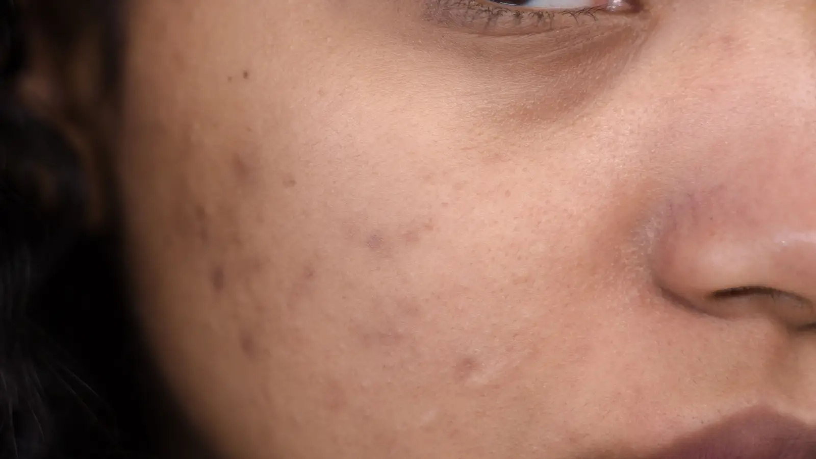
What Dermatologists Do Differently When Microneedling for Dark Spots
Dermatologists treat dark spots with microneedling by operating two levers at once: they set needle depth to control the level of mechanical exfoliation and then exploit the freshly opened microchannels to force melanin-inhibiting agents directly into the skin. Depth is chosen per Fitzpatrick type—because in skin that makes pigment eagerly, the difference between 0.5 mm and 1.0 mm is the difference between clearance and new dark marks. This article lays out the strategic framework that governs those choices, so you can evaluate any protocol against a clinical standard instead of marketing claims.
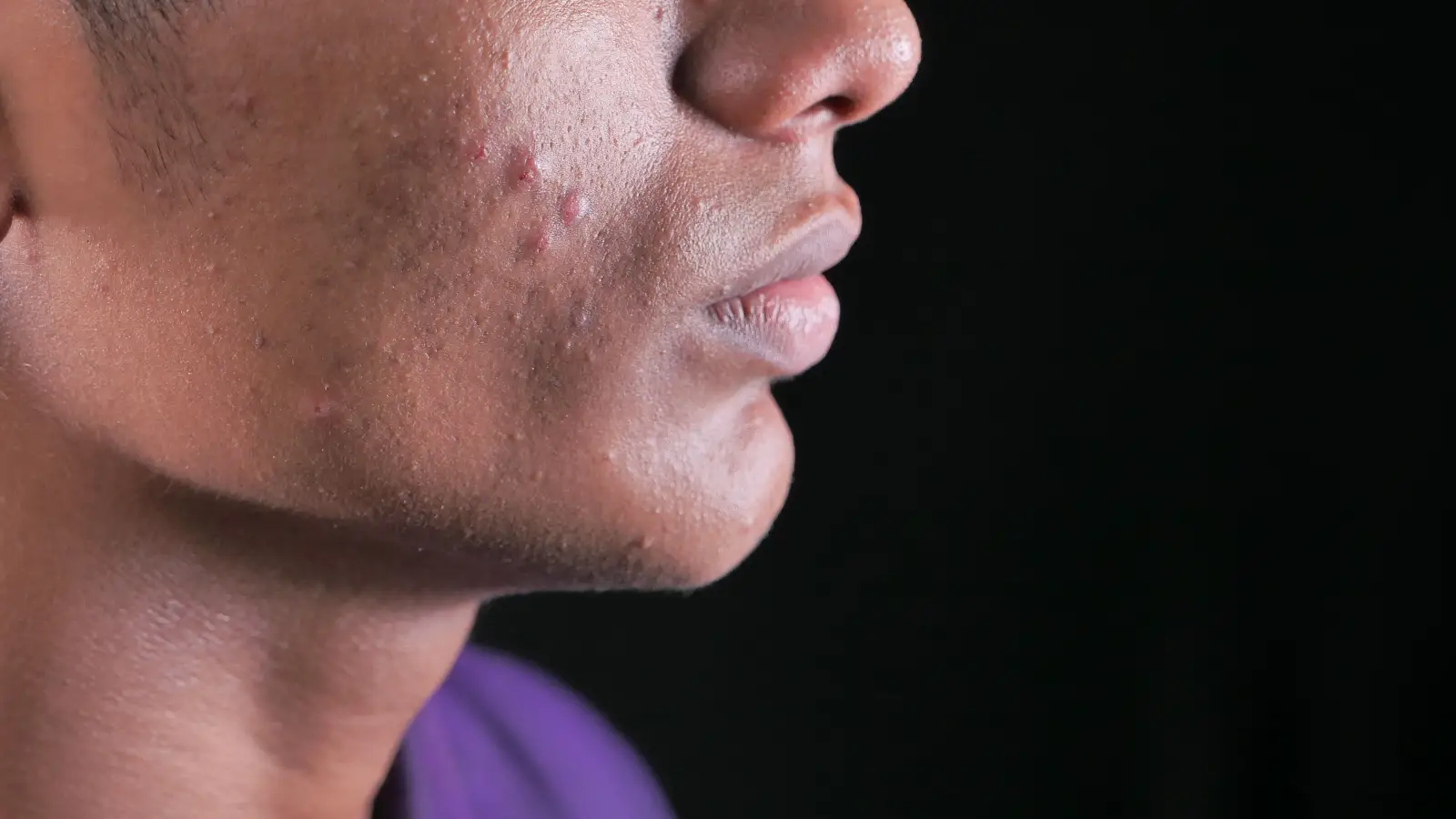
When you understand microneedling for hyperpigmentation as a calibration problem, the nonsense falls away. The treatment’s dual mechanism—physical shedding of keratinocytes loaded with melanin plus a drug-delivery window that boosts penetration of tyrosinase inhibitors tenfold or more—is only half the story. The other half is risk: inflammation from the needle injury itself can ignite melanocytes in Fitzpatrick types III through VI. The dermatologist’s job is to dial in depth, actives, and aftercare so that the exfoliation-plus-delivery effect overpowers the inflammatory drive every time.
The Dual Mechanism That Decides Success or Failure
Microneedling attacks pigment on two fronts. First, the mechanical disruption exfoliates. At 0.25–0.5 mm, needles abrade the outer epidermis, sweeping away melanized cells. This alone yields visible lightening in superficial spots, and because the injury stays above the dermal papillae, it works for any Fitzpatrick type with minimal risk.
Second, and more powerfully, the same punctures create temporary channels that discard the stratum corneum’s barrier function. Actives that would otherwise hover uselessly on the surface now diffuse directly to the basal layer where melanocytes live. Without this delivery, the needle injury can tip the skin into a pigment-producing spiral. With a properly chosen cocktail—tranexamic acid to block melanocyte stimulation, kojic acid to inhibit tyrosinase, niacinamide to stop melanosome transfer—the treatment becomes a net depigmenting event.
Dermatologists exploit this window ruthlessly: they know the microchannels remain patent for only 15–30 minutes, so they flood the field with the active serum during needling, not after, and select agents small enough and stable enough to survive the trip.
Needle Depth and Fitzpatrick Type: The Risk Map
Depth is the master control, and it is never one-size-fits-all. The harder you push into the dermis, the more you remodel—and the more you gamble with post-inflammatory hyperpigmentation in melanin-rich skin.
0.25 mm – 0.5 mm: The Safe Exfoliation Range
At this level, damage stays epidermal. There is no pinpoint bleeding. For Fitzpatrick IV through VI, this is the default start. It exfoliates solar lentigines and post-acne marks while accepting a topical tyrosinase inhibitor without igniting deep inflammation. In-office, dermatologists repeat this shallow protocol every 2–4 weeks, layering the chemical depigmenting effect with each pass.
0.5 mm – 1.0 mm: The Calculated Risk Zone
Now the needles nick the papillary dermis. Fitzpatrick I–III can tolerate this for stubborn mixed dermal-epidermal melasma, provided the actives are on point and sun behavior is airtight. For types IV and V, dermatologists escalate to this depth only after multiple shallow sessions have proven the skin’s melanocytes won’t overreact. Type VI rarely leaves the shallow zone unless the real target is atrophic scars with negligible active pigment.
Above 1.0 mm: Not for Hyperpigmentation
Once needles pass 1.0 mm, they enter the reticular dermis—scar territory, not pigment territory. In darker skin, the inflammatory cascade at this depth routinely outruns any topical suppressant. Dermatologists avoid this range entirely when the goal is dark spot reduction, and if they do use it, it’s paired with a vascular laser or a corticosteroid dampener.
The Agents That Make Microneedling an Anti-Pigment Machine
The active ingredient isn’t an afterthought; it’s the second half of the calibration. Apply the wrong one through microchannels and you’ll get redness, stinging, or paradoxical darkening.
- Tranexamic acid: A small, water-loving molecule that intercepts the plasminogen pathway. Microneedled, it hits melanocytes directly with minimal irritation, making it the anchor of melasma protocols across skin tones.
- Kojic acid: Robs tyrosinase of copper. Effective but prickly; dermatologists buffer it with centella or allantoin to prevent a sting that could be mistaken for PIH.
- L-Ascorbic acid: The low pH required for stability (around 3.5) can inflame microneedled skin. In-office, it’s often swapped for a non-acidic derivative or neutralized quickly so the antioxidant effect lands without the burn.
- Niacinamide: The quiet workhorse. It blocks melanosome transfer to keratinocytes and causes almost no irritation, so it’s safe at any depth and in any Fitzpatrick type.
Dermatologists rarely rely on a single agent. A compounded sterile solution—often tranexamic acid plus niacinamide—is dripped directly onto the skin and worked into the channels with the needling device, using a stamping rather than dragging motion.
In-Office vs. At-Home: What You Can’t Replicate
Consumer microneedling tools fail not because they don’t poke holes, but because they lack the control loop that makes the holes therapeutic rather than destructive.
- Needle hygiene and geometry: In-office cartridges are single-use, medical-grade, and precisely angled so the puncture closes cleanly. Dull, multi-use rollers create micro-tears that amplify inflammation, exactly what hyperpigmented skin doesn’t need.
- Pressure and trajectory: Dermatologists apply perpendicular, even pressure to avoid lateral dragging, which stirs up melanocytes. At-home use tends to be uneven—too much pressure in one spot, too little in another—leading to patchy healing.
- Pre-treatment pause: Retinoids and strong AHAs are stopped a week ahead under a dermatologist’s direction. Over-the-counter rollers rarely come with that instruction, so patients needling over retinized skin multiply their irritant load.
- Post-needling occlusion: A silicone gel or hydrogel sheet applied immediately post-procedure keeps the channels hydrated and maximizes active uptake while calming heat. That step is absent from nearly every at-home routine.
Preventing the Dark Spots Microneedling Can Create
For anyone with melanin-competent skin, post-inflammatory hyperpigmentation is the adversary that must be preempted, not treated after it appears. The protocol starts before the needle.
- Sun defense with iron oxides: A tinted mineral sunscreen that blocks visible light is non-negotiable for the four weeks following treatment. UV and blue light will override any tyrosinase inhibitor.
- Ongoing suppression: Patients apply a melanin-inhibiting cream—azelaic acid or a kojic-niacinamide blend—nightly from the day of the procedure onward.
- Inflammation dampening: For high-risk phototypes, dermatologists often prescribe a mild topical steroid for the first three nights to snuff out the sterile flare before it recruits melanocytes.
- Mechanical rest: No rubbing, no picking, no occlusive liquid makeup, and no heavy sweating for 48 hours. A fresh pillowcase and minimal product contact reduce friction-induced pigment.
These steps are not optional adjuncts; they are the difference between a treatment that lightens and one that darkens. At-home use routinely omits them, which explains why some people walk away with worse pigmentation, not better.
When Microneedling Isn’t the Right Hammer
Microneedling can’t fix every dark mark, and pretending otherwise wastes time and money. Mixed melasma with visible telangiectasias often requires a vascular laser or IPL ahead of needling to collapse the red component, so the brown remnant can be addressed cleanly. Dermal melanocytoses like nevus of Ota sit too deep for any needle to reach; those demand picosecond lasers, and microneedling has no role.
A dermatologist’s real edge here is accurate diagnosis. With a dermatoscope, they separate epidermal pigment from dermal pigment, vascular from melanotic, and inflammatory from stable—then assign the right tool to the right layer. Without that step, you’re treating blindly.
Key Takeaways
- Microneedling works on dark spots via simultaneous mechanical exfoliation and enhanced delivery of melanin inhibitors; both levers must be pulled together.
- Needle depth is the risk dial: 0.25–0.5 mm is safe across all Fitzpatrick types; above 1.0 mm practically guarantees PIH in darker skin and has no pigment-treatment indication.
- The active agents that penetrate the microchannels—tranexamic acid, kojic acid, vitamin C, niacinamide—are chosen not just for efficacy but for irritation potential in each skin type.
- In-office protocols layer sterility, pressure control, pre-treatment prep, and occlusive aftercare into a system that home devices cannot replicate.
- Microneedling is not a universal pigment cure; correct diagnosis of the pigment entity and strategic combination with lasers or peels are part of the dermatologist’s standard operating procedure.
Next Move: Diagnose Before You Needle
The core insight from this framework is that microneedling for dark spots is a calibration exercise. You set depth against the patient’s Fitzpatrick type, select an active cocktail that will tip the net effect toward depigmentation, and wrap the whole thing in a post-procedure regimen that keeps melanocytes asleep. Before booking any treatment—or uncapping any at-home roller—get the specific pigment lesion diagnosed under dermoscopy and ask the provider to explain their depth–Fitzpatrick–active protocol in detail. If they can’t articulate that triad, walk. That’s the difference between a treatment that risks extra pigment and one that removes it.
Pigment Curious
We are people who want to better understand skin pigmentation, dark spots, and how different skin tones respond to skincare and professional treatments. We look beyond simple beauty claims, focusing on scientific evidence, clear explanations, and practical knowledge that helps people make more informed decisions about their skin.