What Derms Verify Before Using Nd:YAG for Dark Spots
A dermatologist does not fire an Nd:YAG laser at a dark spot without first verifying three things: the spot’s depth and melanin composition, the patient’s Fitzpatrick skin type, and the absence of hidden contraindications. No exceptions. The 1064 nm wavelength is the only reason this laser earns its track record on darker skin—it bypasses the melanin‑packed epidermis to target dermal pigment while keeping the surface cool. But that advantage collapses the moment a check is skipped.
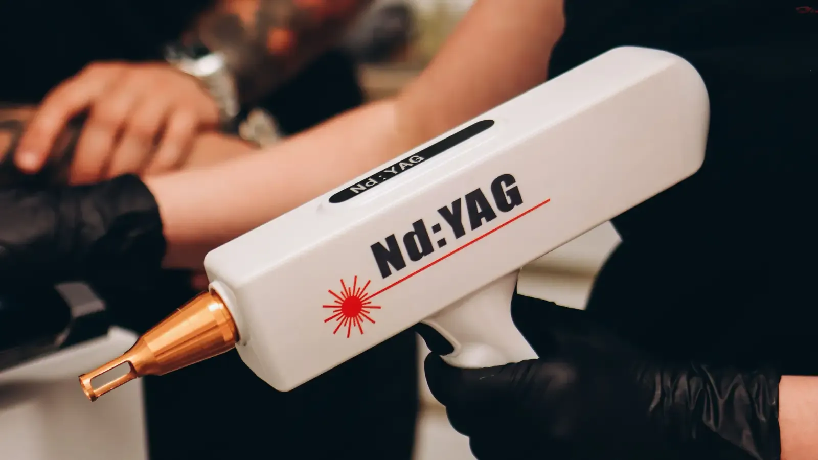
Dermatologists choose the Nd:YAG over shorter‑wavelength lasers because the 1064 nm beam has a fundamentally lower melanin absorption coefficient—meaning it heats the epidermis less and penetrates deeper. That’s the physics. But physics alone doesn’t guarantee a safe treatment. The selection process is a clinical gauntlet: a step‑by‑step verification that determines whether the spot is a genuine candidate for deep pigment targeting. Until every check clears, the laser stays off.
The Three Dimensions That Decide Nd:YAG’s Role
Every Nd:YAG treatment decision turns on three hard conditions: what the spot is made of, what the patient’s skin will allow, and how the laser’s settings can be tuned to keep both safe. Skip any one of these and the procedure becomes guesswork.
Melanin biology is the first condition. Spots live either in the epidermis, the dermis, or both. The Nd:YAG’s 1064 nm beam is absorbed by melanin, but only weakly compared with shorter wavelengths—that’s why it penetrates deep before heating pigment. For dermal collections (like many PIH marks and nevus of Ota), this is exactly what’s needed. For purely superficial spots, a 532 nm or IPL is the sharper tool. The dermatologist’s first job is to locate the pigment precisely. Anything less is a shot in the dark.
The second condition—and the one that separates the Nd:YAG from nearly every other laser—is Fitzpatrick phototype. Darker skin (IV–VI) holds larger, more excitable melanosomes. A wavelength that barely warms light skin can torch an epidermis rich in melanin. The 1064 nm wavelength opens a narrow therapeutic window where dermal pigment can be destroyed while the epidermis stays below injury threshold. But that window slams shut the instant skin type is read incorrectly or PIH history ignored. A dermatologist verifies not just the Fitzpatrick number, but the skin’s real‑world behavior: does it darken after a scratch, a peel, a pimple? That history is the true safety gauge.
The third condition is the laser’s configurability. Fluence, spot size, pulse duration, cooling—each parameter must be dialed in for the specific skin type and lesion depth. On darker skin, fluence is driven low—typically 3–8 J/cm²—and a test spot is non‑negotiable. Nothing is copied from a manual. The dermatologist custom‑builds the protocol from the ground up.
First: Spot Depth and Melanin Characterization
The first clinical verification is a hard diagnosis. “Dark spot” is not a diagnosis. Dermatologists separate lentigines from flat seborrheic keratoses, PIH from melasma, dermal nevi from café‑au‑lait macules. Everything that follows depends on getting this right.
A dermoscope reveals whether the pigment sits in the epidermis (network‑like, regular) or deeper, with a bluish‑grey cast. A Wood’s lamp reinforces the finding: epidermal pigment lights up; dermal pigment stays dark. If a lesion shows any asymmetry, rapid change, or atypical features, a biopsy rushes to the front of the queue—lasers never touch an un‑biopsied suspicious spot.
Why this verification comes first is simple physics. The Nd:YAG’s 1064 nm beam is built for depth. Fire it at a superficial lentigo and the energy sails right past the target, heating the dermis with no clearance. Worse, if the surface lesion is actually melasma, the deep heat stokes inflammation and triggers rebound darkening weeks later. Lesion depth is the fork in the road. The dermatologist uses it to rule out what shouldn’t be treated with 1064 nm at all.
Done well: the spot is definitively classified as a dermal or mixed melanin lesion that genuinely needs 1064 nm penetration, malignancy is ruled out, and the baseline is photographed.
Second: Fitzpatrick Skin Type and PIH Risk Assessment
Skin type verification is not a box‑tick. It’s a risk‑prediction exercise. A dermatologist maps the patient’s PIH history—every past insult, every time a pimple left a shadow, every instance of darkening after a scrape or peel. That data outweighs the Fitzpatrick number alone. A type IV skin that never hyperpigments is a safer canvas than a type III that does.
The Nd:YAG’s safety advantage is real, but it’s conditional. In darker phototypes, PIH rates hover around 2–5% even with 1064 nm when fluences aren’t pulled back enough. So the dermatologist delivers a blunt risk‑benefit statement: the treatment offers clearance, but it carries a small chance of temporary darkening that demands sun avoidance and rigorous sunscreen adherence for weeks. If the patient won’t commit to that sunscreen discipline, the laser stays unused. No negotiation.
This verification delivers a personalized safety margin that directly controls parameter selection next. Without it, the laser is flying blind.
Third: Contraindication Screening—What Rules Out Nd:YAG Immediately
Before any pulse, the dermatologist runs a hard stop: conditions that make Nd:YAG unsafe must be eliminated first.
Absolute stops include active infection, a photosensitivity disorder (lupus, porphyria), recent isotretinoin use (within 6–12 months), and suspicious lesions not yet biopsied. Pregnancy is a stop—not because of known fetal risk, but because hormonal hyperpigmentation becomes unpredictable and safety data doesn’t exist.
Relative stops demand caution, not cancellation. Melasma is the biggest. Nd:YAG can exacerbate epidermal melasma if applied like a standard spot removal. A recent tan or sunburn primes the epidermis with melanin and invites burns. Photosensitizing medications—tetracyclines, amiodarone, St. John’s Wort—and even topical retinoids that thin the skin all raise the stakes. The dermatologist interrogates the medication list and supplement cabinet.
Melasma identification is a verdict point. If the spot is actually melasma—bilateral, blotchy, hormonally driven—a standard Q‑switched Nd:YAG approach will likely provoke rebound hyperpigmentation worse than the original mark. Low‑fluence “laser toning” might be an option, but only as part of a topical‑first strategy. When melasma is on the table, full‑energy spot removal is off.
Fourth: Parameter Selection and the Test Spot
With the lesion confirmed and the skin’s tolerance mapped, the dermatologist now builds the laser protocol from scratch.
Key parameters: fluence (3–8 J/cm² for darker skin, 5–10 J/cm² for lighter types), spot size (2–4 mm), pulse duration (nanosecond Q‑switched for pigment), repetition rate, and cooling method. The selection is a custom fit—no part is lifted from a generic chart.
The test spot is the gatekeeper. A small, hidden area is treated with the planned settings and watched for two to four weeks. If it heals flat, without blistering, crusting, or PIH, the full treatment can proceed. If even mild PIH creeps in, the dermatologist pulls back the fluence, widens the spot size, or reduces passes. The test spot isn’t optional—it’s the only real‑world proof that the chosen parameters work on this specific patient’s skin.
Done well means the protocol was designed for the individual and the test spot confirmed it.
How One Patient’s Workup Followed All Four Steps
Consider a Fitzpatrick V man with a single dark, bluish‑grey cheek macule left behind after cystic acne. The spot hasn’t budged in months.
First verification: dermoscopy shows smooth, grey‑blue pigment—classic dermal melanophages. Wood’s lamp: no enhancement. Depth confirmed as dermal PIH. No suspicious features. This is a textbook Nd:YAG target.
Second: PIH history check. The patient darkens predictably after every bug bite and razor bump; a previous glycolic peel needed a full year to normalize. Risk is high. The dermatologist explains the 1064 nm safety advantage but insists on low‑fluence parameters and strict SPF 50+ sunscreen starting two weeks pre‑treatment. The patient commits.
Third: no medications, no isotretinoin, no recent sun. The spot is clearly PIH, not melasma. Contraindications are absent.
Fourth: Q‑switched Nd:YAG, 3 mm spot, fluence at 3.5 J/cm² (half the typical for lighter skin), with cryogen cooling. A test spot is placed behind the jawline. Three weeks later, it’s calm, slightly lighter, with zero PIH. Treatment proceeds. Clearance follows over two sessions.
When Verification Steps Are Skipped—and What It Costs
Shortcuts extract a price. These three failures repeat most often.
Treating a Superficial Spot as if It Were Deep
Assuming a dark spot is dermal because it looks dark is a depth‑blind mistake. A 1064 nm pulse fired at a purely epidermal lentigo sails past the target, wastes the session, and heats the dermis for nothing. A 532 nm or cryotherapy would have cleared it. No depth check, no clearance.
Using Nd:YAG as if Skin Type Doesn’t Matter
The Nd:YAG is safest for dark skin—but “safest” isn’t “safe regardless.” If a dermatologist applies fluences meant for Fitzpatrick II onto type V skin, blistering, PIH, or hypopigmentation are the predictable result. Skin type verification isn’t just a number; it’s the entire permission structure for the energy setting. Skip it, and the laser becomes a pigment factory instead of a eraser.
Failing to Identify Melasma
Melasma mimics other spots. Treat it with a standard Q‑switched Nd:YAG and the hormonal pigmentation engine revs up, producing a rebound dark mark worse than the original. Recognizing melasma before the laser ever touches the skin is the difference between improvement and months of damage control. When this step fails, the recovery often drags on for a year or longer.
Quick Reference
| Verification Stage | Primary Goal | Key Action |
|---|---|---|
| Spot depth and melanin type | Confirm dermal pigment and rule out superficial-only lesions | Dermoscopy, Wood’s lamp, biopsy if suspicious |
| Fitzpatrick skin type & PIH history | Estimate risk of pigmentary side effects | Detailed PIH history, patient counseling, risk discussion |
| Contraindication screening | Eliminate conditions that make Nd:YAG unsafe | Medical history, medication review, melasma identification |
| Test spot and parameter tuning | Validate safety and efficacy in that individual | Conservative test parameters, 2–4 week observation, adjust if needed |
FAQ
Is the Nd:YAG laser safe for dark skin?
Yes, when used correctly. The 1064 nm wavelength gives Nd:YAG the lowest risk of pigment alteration among all lasers for skin types IV through VI. But safety is not automatic—it depends on conservative parameters, a test spot, and strict pre‑treatment verification. Cut corners and you invite PIH.
Can Nd:YAG treat all types of dark spots?
No. Nd:YAG works best on dermal or mixed dermal‑epidermal pigment, such as deep PIH, nevus of Ota, and some flat seborrheic keratoses. Superficial epidermal spots like freckles or thin lentigines often respond better to shorter‑wavelength lasers or IPL. Melasma requires a specialized, low‑energy approach and is not a straightforward Nd:YAG indication.
How can I tell if my dark spot is suitable for Nd:YAG before seeing a dermatologist?
You can’t determine this at home. Depth assessment requires dermoscopy and Wood’s lamp examination by a trained clinician. Spots that are raised, rapidly changing, or painful should be evaluated urgently to rule out skin cancer. A dermatologist consultation is the only reliable way to confirm Nd:YAG candidacy.
What should I do if I develop PIH after Nd:YAG treatment?
Stop further laser sessions immediately and return to your dermatologist. PIH management usually involves topical depigmenting agents (hydroquinone, azelaic acid, kojic acid), strict sunscreen, and sometimes low‑dose topical corticosteroids. Most PIH fades over months, but additional lasers will worsen it if applied before the skin stabilizes.
Does the Nd:YAG laser hurt?
Most patients describe Q‑switched Nd:YAG pulses as a quick rubber‑band snap with mild prickling. Cooling reduces sensation to a tolerable level. For small areas, topical anesthesia isn’t needed. Long‑pulsed Nd:YAG feels warmer but is typically less sharp.
From Verification to Safe Treatment
The Nd:YAG laser clears dermal dark spots on darker skin only when the clinician moves through a defined sequence: diagnose spot depth, assess PIH risk, screen for contraindications, and test the parameters on a small patch. Nothing else matters if those steps break. Start with depth—if the spot hasn’t been examined under a dermatoscope to confirm it’s dermal, that’s the first action to take with a board‑certified dermatologist.
Pigment Curious
We are people who want to better understand skin pigmentation, dark spots, and how different skin tones respond to skincare and professional treatments. We look beyond simple beauty claims, focusing on scientific evidence, clear explanations, and practical knowledge that helps people make more informed decisions about their skin.
This is the most thorough explanation I’ve seen about the pre-treatment checks. I have a dark spot on my jawline and now feel much more prepared to ask the right questions at my consultation. Thanks for laying it out step by step.
I had Q-switched Nd:YAG on a deep PIH mark (I’m Fitzpatrick IV) and the test spot was a game changer. My derm used super low fluence and it took two sessions but the mark’s almost gone. You’re so right about the sunscreen discipline—I wore SPF 50 every single day for a month.
I have a brown spot on my forehead that my derm said is melasma, and it gets darker every summer. Topicals haven’t helped much. Could a very low-fluence Nd:YAG still be tried, or is that just too risky?
That’s an excellent question. Low-fluence Nd:YAG can be an option for melasma, but only as part of a careful, often topical-first strategy. Because melasma can easily rebound, any laser trial should start with a very conservative test spot and weeks of observation. It’s essential to work with a board-certified dermatologist who has experience managing melasma with lasers—not all do.
Really helpful article, saving this for my next derm appointment.