Why Your ‘Age Spot’ Is Probably Not an Age Spot
That flat brown patch you call an ‘age spot’ is, in consistent dermatologic terms, a solar lentigo — a macule produced by cumulative ultraviolet radiation, not by the passage of time. Calling it an ‘age spot’ obscures the real cause. The following material identifies solar lentigines, sun spots, and age spots as one and the same lesion, then provides reproducible criteria — border definition, body distribution, and sun‑exposure history — to separate them from ephelides (freckles) and flat seborrheic keratoses.
A systematic approach matters because each entity follows a different trajectory and carries distinct monitoring requirements. The decision‑tree that follows does not replace clinical dermoscopy, but it anchors self‑assessment in observable, measurable features.
Quick Spot Assessment
| If Your Spot… | It’s Most Likely… | Because… |
|---|---|---|
| Appeared after your 30s on sun‑exposed skin and does not fade | Solar lentigo (age spot) | Chronic sun exposure triggers epidermal melanin accumulation; no seasonal fluctuation |
| Has been present since childhood, lightens in winter, darkens every summer | Ephelides (freckles) | Genetically determined, sun‑triggered but reversible; fades without UV |
| Has a slightly waxy feel, a sharp border, and looks as if it could be scraped off | Flat seborrheic keratosis | Benign epidermal proliferation, not directly sun‑related; often rough or stuck‑on |
When the Spot Persists Year‑Round on Sun‑Exposed Skin
The lesion that remains unchanged from January to August on the cheeks, dorsal hands, shoulders, or upper chest is, in nearly every case, a solar lentigo. Cumulative UV radiation drives melanocytes to deposit pigment into keratinocytes, creating a flat macule that measures from a few millimeters to over a centimeter. Its color spans light tan to dark brown, and its margins are distinct but not scalpel‑sharp.
Interpreting this finding as a solar lentigo means you accept that it signals a history of photoaging. The practical cost is that you may fixate on one prominent spot while overlooking adjacent, smaller lentigines and the background dermatoheliosis that a dermatologist would register under polarized dermoscopy as a uniform, reticulated pigment network.
When the Spot Fades Every Winter
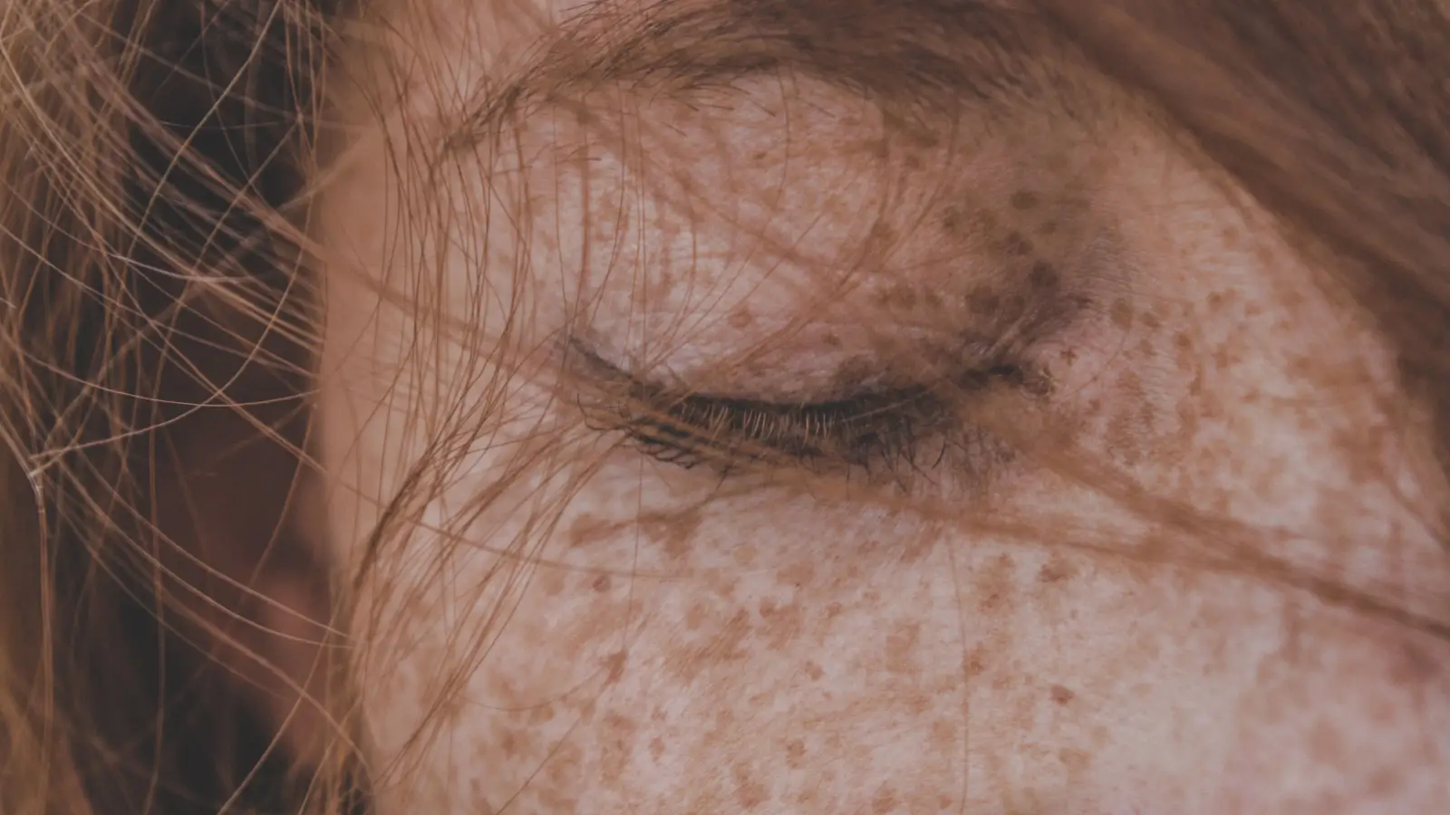
A brown mark that nearly vanishes during low‑UV months and reappears each summer follows the pattern of an ephelid — a freckle. These are genetically programmed, most common in Fitzpatrick skin types I and II, and cluster on the face, forearms, and upper back. Histologically, they show basal‑layer hyperpigmentation without an increase in melanocyte count, a clear difference from a solar lentigo.
The benefit of identifying this pattern is reassurance: ephelides are benign and often fade with sun avoidance. The risk is that a freckle field can camouflage an outlier — a lentigo maligna or a dysplastic nevus — especially if one spot refuses to fade in winter. That single stubborn lesion violates the seasonal rule and requires dermoscopic scrutiny.
When the Spot Feels Waxy or Stuck‑On
A flat brown lesion that, on close inspection, has a greasy surface texture and a sharply demarcated, almost pasted‑on border is unlikely to be a solar lentigo. The description fits a flat seborrheic keratosis. These common benign growths appear on both sun‑exposed and sun‑protected skin and often show a subtle elevation detectable with the fingertip, even when they look flat to the naked eye. A magnifying mirror may reveal tiny keratin cysts or comedo‑like openings.
Correctly categorizing the spot as a seborrheic keratosis removes one source of anxiety. The caution is that certain melanomas — particularly verrucous variants — present as seborrheic‑keratosis‑like lesions. The reliable differentiator is a dermoscopic pattern that is orderly in a seborrheic keratosis and disordered in melanoma. When in doubt, the spot deserves an in‑person examination.
Stop Weighing the Names: Solar Lentigo, Age Spot, and Sun Spot Are One Lesion
The diagnostic terms solar lentigo, age spot, sun spot, and liver spot all designate the same acquired melanotic macule. ‘Liver spot’ is a historical misnomer with no connection to hepatic function. The variation in names adds no clinical value. Focus instead on the features that actually separate a solar lentigo from a freckle or a seborrheic keratosis: border regularity, color uniformity, symmetry, and seasonal behavior.
Specific Features That Rule Out a Solar Lentigo
A solar lentigo is defined by three hard constraints: it must be completely flat (macular), it must arise on chronically sun‑exposed skin, and it must evolve only minimally over years. Any deviation from these constraints directs the differential diagnosis elsewhere.
- Palpable texture: Even a faint roughness felt with the fingertip disqualifies a pure solar lentigo. Elevation points toward a seborrheic keratosis, an actinic keratosis, or another pathology that may require treatment.
- Location outside sun‑exposed zones: A brown spot on the lower back, buttocks, genitals, or oral mucosa is not a solar lentigo. Such sites demand an alternative explanation.
- Irregular morphology: Asymmetry, multiple colors (tan, black, pink, gray) within one spot, or a border that is notched rather than smoothly defined excludes a simple lentigo. These are ABCDE criteria for melanoma, and dermoscopy is urgently indicated.
- Accelerated change: A solar lentigo changes imperceptibly over years. A spot that darkens, enlarges, or bleeds over weeks or months is not a solar lentigo. Timely professional evaluation is the only safe response.
A dermatologist’s dermoscopic examination remains the reference standard. The criteria here are designed to help you determine when that standard is mandatory.
Frequently Asked Questions
Are ‘age spot’ and ‘liver spot’ the same thing?
Yes. Both are colloquial labels for a solar lentigo. Neither reflects any internal organ dysfunction; the lesion is confined to the skin and caused by sun exposure.
Can a solar lentigo become cancerous?
A solar lentigo itself does not have malignant potential. However, it serves as a clinical sign of significant cumulative UV damage, which raises the risk of melanoma, basal cell carcinoma, and squamous cell carcinoma elsewhere on the skin. A solar lentigo that changes in size, color, or shape must be examined to exclude lentigo maligna melanoma.
How can I reliably tell a freckle from a solar lentigo without a dermatoscope?
Observe the spot for one full year. A freckle will visibly fade in winter and intensify in summer. A solar lentigo will remain stable. Freckles also tend to be smaller, lighter, and more numerous, forming confluent patches on sun‑exposed areas.
Will a solar lentigo disappear if I stay out of the sun?
No. Unlike ephelides, solar lentigines do not regress with sun avoidance. They are permanent unless removed by a targeted intervention such as a topical bleaching agent, laser, or cryotherapy.
What to Do Next
For the most common scenario — a flat brown spot on sun‑stressed skin, appearing after early adulthood and persisting year‑round — the most probable diagnosis is a solar lentigo. The single deciding question is: “Does this spot hold its appearance across all seasons, or does it come and go?”. A stable, unchanging spot favors solar lentigo. Any ambiguity in the answer is a direct prompt to see a dermatologist.
Pigment Curious
We are people who want to better understand skin pigmentation, dark spots, and how different skin tones respond to skincare and professional treatments. We look beyond simple beauty claims, focusing on scientific evidence, clear explanations, and practical knowledge that helps people make more informed decisions about their skin.