3 Pigment Depths That Decide Your Spot Treatment Success
The diagnostic problem behind every stubborn dark spot is fundamentally a question of depth. Excess melanin either sits in the epidermis, the dermis, or straddles both layers simultaneously. Where it sits physically determines which chemical or energy-based tool can reach it; ignore that constraint and you’re not treating, you’re gambling. The clues that make the depth readable without a Wood’s lamp are already visible: the spot’s dominant color in natural light and the crispness of its boundary. Brown and sharply outlined points to superficial epidermal pigment. Blue-gray and smudgy points to deep dermal pigment. Mixed spots show both signatures in different zones. The payoff for reading these signs correctly is the difference between choosing a topical that actually has access to its target, a peel that works, or a laser that can deliver energy to the right layer without triggering more pigment production in melanin-reactive skin.
One Cheekbone Mark That Became a Depth Proposition
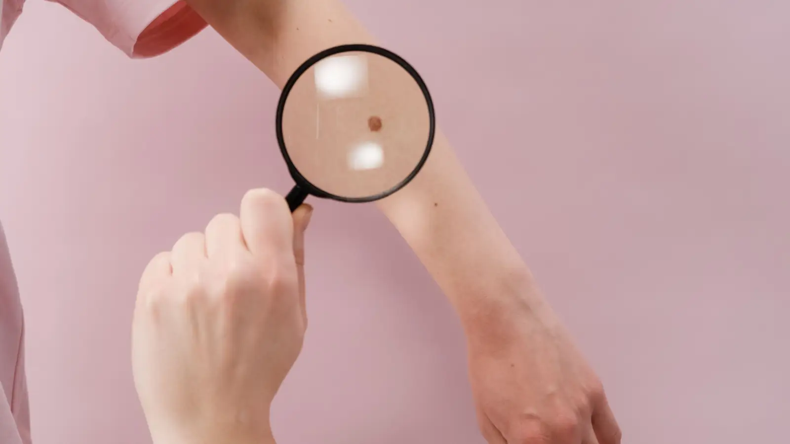
A 34-year-old woman with medium-brown skin watches a persistent mark on her left cheekbone after a modest acne bump finishes healing. For six weeks she applies a 2% hydroquinone cream paired with a vitamin C serum. The outer rim of the spot obediently fades into a faint tan halo—almost invisible now. The center, however, stays put as a dusky, blue-gray smudge, refusing to budge an inch. The mark isn’t one lesion; it’s two different pigment populations living at two different depths, and the differential response to the same cream is the proof. This split response turns a routine treatment attempt into a practical diagnostic tool: the part that faded behaves like an epidermal spot, and the part that didn’t behaves like a dermal one. From that point on, any plan that treats both zones identically will be wrong by design.
Depth Signatures: What Color and Border Blur Are Actually Reporting
The visual characteristics of a pigment spot—its hue and its edge quality—correlate with layer location in a predictable way, because light interacts with superficial melanin and deep melanin according to different scattering physics.
The Epidermal Signature: Brown, Bordered, and Vulnerable to Topicals
Melanin confined to the epidermis absorbs and reflects light without the interference of deep-tissue scatter, producing a brown color that stays within a tight, well-defined edge. The pigment’s proximity to the skin surface makes it accessible to topical tyrosinase inhibitors like hydroquinone, kojic acid, and azelaic acid, as well as to superficial chemical peels that remove only the stratum corneum and the uppermost living epidermis (glycolic acid at 30–50% or salicylic acid at 20–30% for oily skin types). The fading outer halo on the cheekbone conformed to this pattern exactly: it lightened with a standard serums-and-exfoliation routine over a matter of weeks. For pure epidermal pigment, the treatment logic is linear—suppress new melanin synthesis, accelerate surface shedding, and block UV-driven reactivation.
The Dermal Signature: Blue-Gray, Blur-Stricken, and Requiring Selective Photothermolysis
Deeper melanin lodged among collagen bundles in the dermis scatters blue light preferentially (the Tyndall effect), giving the spot a blue, gray, or faintly purple appearance. The border appears hazy because the pigment is dispersed in a three-dimensional volume rather than packed into a flat epidermal plane. Topical agents barely diffuse to the dermis in meaningful concentrations, and superficial peels are structurally incapable of reaching that depth—they end at the epidermal-dermal junction. The central smudge on the cheekbone demonstrated this limitation silently; the melanin there didn’t “resist” the cream, it simply never encountered it. The tool with the right reach is a Q-switched laser, most commonly the 1064 nm Nd:YAG, which uses nanosecond pulses to fracture dermal melanin while sparing the overlying epidermis enough to keep the risk of post-inflammatory hyperpigmentation manageable on melanin-rich skin. Low fluence and a large spot size are not optional refinements in this context; they are what prevents the treatment from turning into a new hyperpigmentation episode by overheating the basal layer. Test spots remain a required step, because the margin between therapeutic energy and pigment-induction energy can be narrow.
The Mixed-Depth Profile: Why a Spot Clears Only Halfway and Stalls
Most stubborn pigment marks are a composite: an epidermal layer formed by superficial inflammation, plus a dermal deposit that results from deeper trauma or prolonged melanocyte stimulation. The cheekbone’s dual behavior—outer ring fading, inner core persisting—is the standard presentation of a mixed-depth spot. The clinical danger is misreading the partial improvement as a sign that the treatment pathway is correct and simply needs more time or more intensity. That logical error leads to over-exfoliation, stronger acid peels, or a high-fluence laser pass, all of which can aggravate the dermal component and deposit even more pigment. A more rigorous strategy sequences the interventions: first resolve the epidermal layer with a tyrosinase inhibitor and disciplined photoprotection for about eight to twelve weeks, then reassess. If a dermal residue remains, a low-fluence Q-switched laser becomes a rational next step, not a reaction to failure. The sequence keeps inflammation—and the consequent melanocyte activity—at its lowest possible level throughout.
Situations Where Depth Ceases to Be Theoretical
Depth isn’t an academic curiosity; it’s the pivot point in several common hyperpigmentation presentations.
Post-inflammatory hyperpigmentation (PIH) following acne
Inflamed acne lesions that breach the dermal-epidermal junction often deposit pigment in both layers. The resulting mark may show a superficial brown cast that lightens within weeks and a deeper wash that lingers for months. The border blur at the perimeter of the remaining pigment can tell you whether the dermal component is significant—hazy edges lean dermal, crisp edges lean epidermal—and therefore whether a topical-only plan is realistic.
Melasma on the cheeks and forehead
Melasma is a mixed-depth condition by default in many cases, driven by vascular and hormonal pathways that pump melanin into both the epidermis and dermis. This explains why a chemical peel can temporarily erase the surface component only for the pattern to reemerge as the dermal melanin casts its shadow again. Treatment decisions here are especially consequential: an epidermal-dominant melasma may tolerate a mild glycolic peel, but a dermal-dominant presentation should steer the conversation away from aggressive modalities and toward sustained pigment suppression, because even low-fluence lasers risk rebound hyperpigmentation on reactive skin.
Solar lentigo on sun-exposed forearms
A solar lentigo tends to be a flat, uniformly brown spot with a sharp border, indicating entirely epidermal pigment. These respond reliably to cryotherapy, light chemical peels, or simply consistent sunscreen and a pigment-inhibiting serum. Their predictive ease makes them useful for calibrating your eye to the color-and-border reading process before tackling trickier mixed lesions.
The Single Most Costly Assumption About Dark Spots
Treating every pigmented spot as if it were a surface stain that any brightening serum can erase is the assumption that sinks the most treatment plans. The cheekbone mark rejected that assumption in clear terms: the outer rim responded, the inner core did not. The failure wasn’t the product’s efficacy; the failure was asking a topical to act on melanin it couldn’t reach. The collateral damage comes when someone interprets the stalled progress as a cue to escalate—deeper peels, microneedling, or an off-label laser pass with the wrong wavelength—triggering melanocytes that were previously quiet. On skin that hyperpigments easily, the spot can return larger and more stubborn than before. The corrective mental model is to treat depth as the primary sorting variable: brown and crisp → treat it like a surface target; blue-gray and blurry → treat it like a deep target, and accept that surface tools have a hard physical limit.
Questions That Follow a Depth-Aware View of Pigment
How can I tell dermal pigment from epidermal pigment without a Wood’s lamp?
Stretch the skin gently. An epidermal spot stays brown and maintains a sharp border. A dermal spot remains blue-gray and its edges stay indistinct. Ambient daylight, rather than specialized equipment, is all that’s required.
If a spot looks blue or gray, is it always dermal melanin?
Not exclusively. Hemosiderin deposition from older vascular injury or deep bruising can produce similar cool tones, but those usually fade over weeks to months. Persistent, non-blanching blue-gray marks that follow acne or trauma are more likely dermal melanin and should be assessed for laser candidacy.
Which laser parameters can treat dermal hyperpigmentation on darker skin without causing new spots?
A Q-switched 1064 nm Nd:YAG with a large spot size (6–8 mm) and deliberately low fluence—starting near the threshold at which pigment shattering occurs without visible epidermal whitening—is the commonly referenced starting point. The 1064 nm wavelength penetrates deeply while minimizing absorption by epidermal melanin. Still, even optimized parameters cannot eliminate risk entirely; a small, inconspicuous test spot and a multi-week observation period between sessions are necessary precautions, not optional courtesies.
Can a medium-depth or deep chemical peel reach dermal pigment?
Deep peels can reach the papillary dermis, but they do so by inducing controlled injury, which on hyperpigmentation-prone skin routinely triggers post-inflammatory hyperpigmentation that is worse than the original spot. Reaching the depth doesn’t equal treating it safely. A laser with wavelength selectivity targeting melanin offers a more precise path with less collateral pigment risk when dermal clearance is the goal.
The Shortest Possible Memory Hook
The cheekbone mark distilled the entire decision tree into two visual checks. Brown and crisp means the melanin is close to the surface, where creams and gentle peels can work. Blue-gray and blurry means it’s sitting in the dermis, beyond the physical reach of anything applied topically—and a treatment that can’t bridge that gap won’t close it.
Pigment Curious
We are people who want to better understand skin pigmentation, dark spots, and how different skin tones respond to skincare and professional treatments. We look beyond simple beauty claims, focusing on scientific evidence, clear explanations, and practical knowledge that helps people make more informed decisions about their skin.