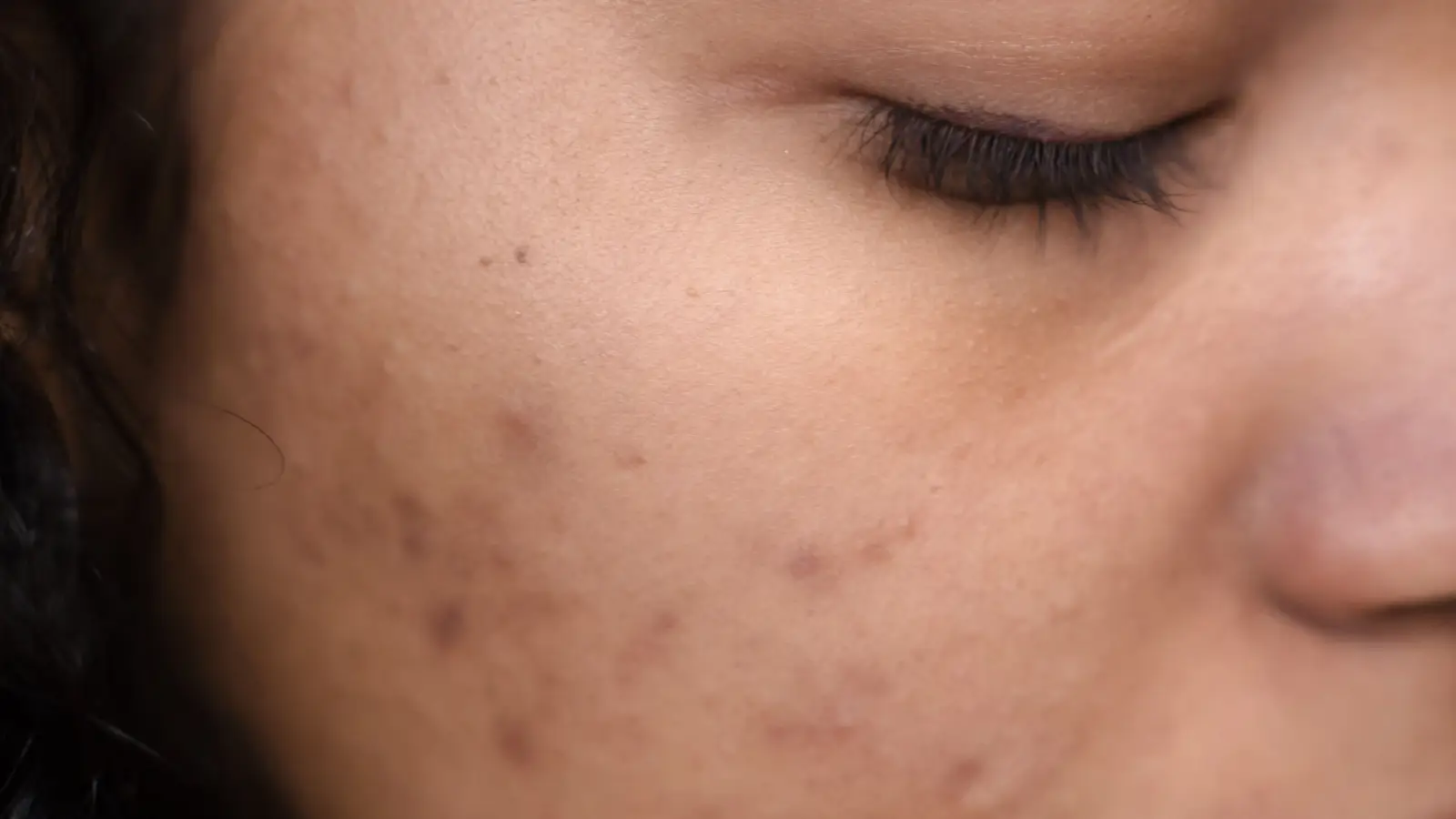
Fade Post-Inflammatory Hyperpigmentation When Nothing Works
The dark marks from a past breakout or rash haven’t budged despite trying brightening serums, vitamin C, and drugstore chemical exfoliants. This persistence usually signals a mismatch between the treatment’s reach and the pigment’s actual location, compounded by a lack of adjustment for skin’s melanocyte reactivity. By the end of this guide, you will have a clear, stepwise method to determine whether the pigment is epidermal or dermal, and how to select interventions that safely work with your skin tone — not against it.
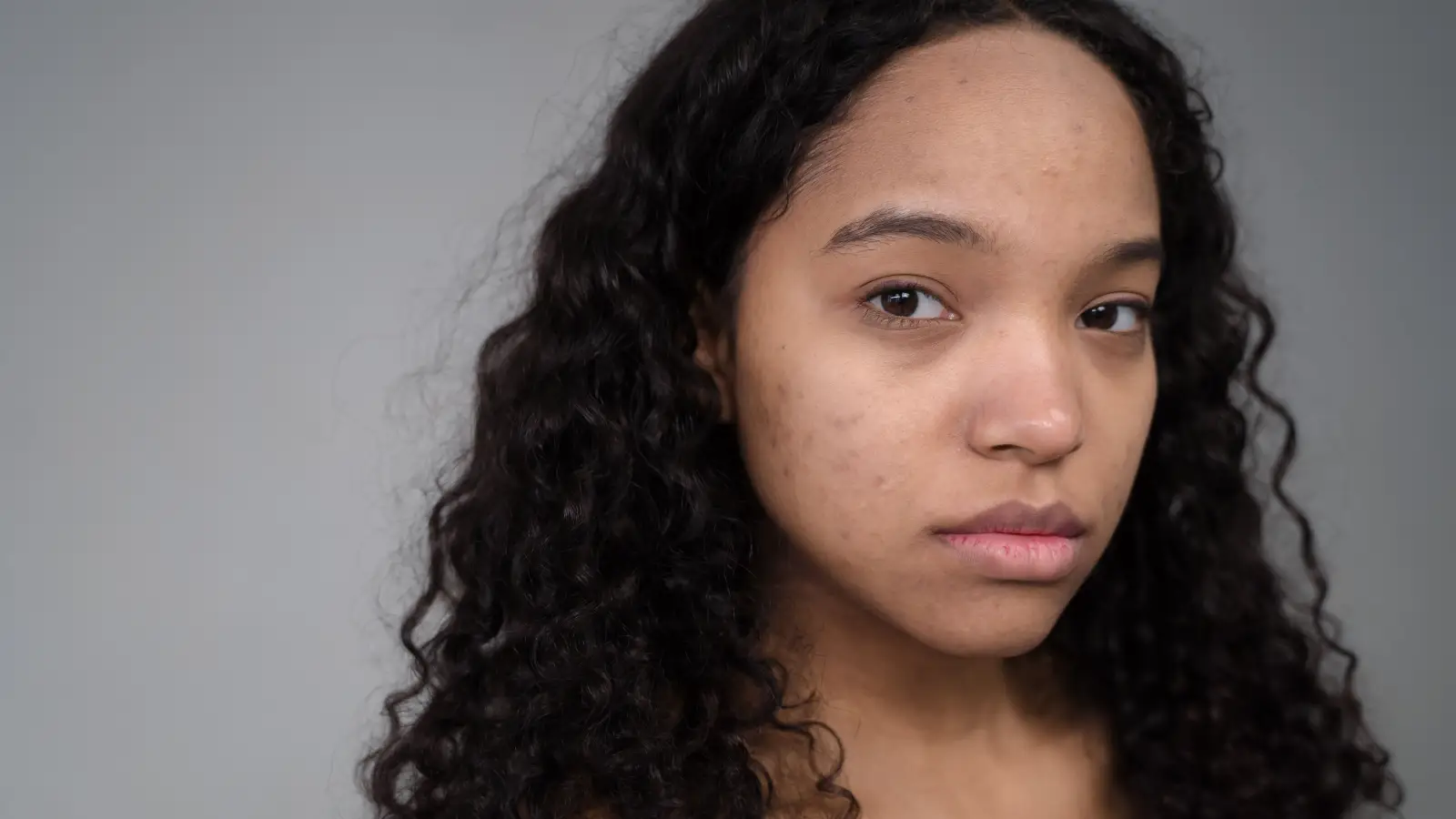
Why Routine Treatments Fail Against Stubborn Post-Inflammatory Hyperpigmentation
Post-inflammatory hyperpigmentation originates when inflammation from acne, eczema, burns, insect bites, or harsh skincare prompts an overproduction and uneven release of melanin. The melanocyte cells at the base of the epidermis react to inflammatory mediators, primarily prostaglandins and cytokines, by ramping up melanin synthesis. In lighter skin types, the pigment typically deposits in the upper epidermal layers and fades along with natural skin renewal. In skin of color, the same inflammatory event often forces melanin into the deeper dermis, where it becomes trapped among collagen fibers and is cleared very slowly, if at all, by surface exfoliation.
When nothing has worked, the core problem is frequently depth-related. Over-the-counter brighteners like kojic acid or diluted vitamin C esters inhibit tyrosinase at the outermost living cell layer. They cannot dislodge pigment that has settled below the basement membrane. A second common reason is that repeated application of irritant exfoliants sustains a low-grade inflammation that keeps melanocytes active, so the marks darken rather than fade. Persistent PIH thus falls into one of three categories: predominantly dermal pigment, ongoing subclinical irritation, or a treatment intensity that triggers rebound melanogenesis on reactive skin.
The Two Depths of PIH: Epidermal Versus Dermal Pigment
Treatment selection fails without first establishing depth. This is the logical starting point before any product or procedure is introduced.
Identifying Epidermal PIH Without a Clinic Visit
Epidermal hyperpigmentation sits within the surface layer that continuously sheds. The marks appear brown to tan with crisp edges. When the skin is stretched gently, the spot often lightens because the pigmented keratinocytes are pulled apart. These lesions respond to tyrosinase inhibitors, superficial exfoliants, and sun protection. Without ongoing inflammation, they typically fade within 6 to 12 months on their own.
Pinpointing Dermal and Mixed PIH
Dermal pigmentation occupies the collagen-dense dermis. The spots have a greyish-blue, ashy, or violet tint — the Tyndall effect scattering light off deep melanin. Stretching does not alter their appearance. The body’s macrophages engulf dermal melanin but degrade it slowly; complete clearance can take years, and spontaneous resolution is rare. Many individuals present with mixed PIH, a brown superficial layer over a persistent grey undercast. This explains why a cream that lightens the surface leaves an unfaded shadow behind: the deeper melanin was never reached.
In clinic, a Wood’s lamp examination or dermoscopy will differentiate the planes in under a minute. This exam avoids months of wasted effort on wrong-layer treatments.
How Skin Tone Dicts Treatment Risk and Outcomes
Skin tone, categorized by Fitzpatrick phototype, shapes melanocyte biology in ways that directly affect the safety of any intervention. An energy level or peel concentration tolerated by phototype I–II without incident can produce new, darker PIH in phototype IV–VI.
The Histology Behind Melanocyte Reactivity
In lightly pigmented skin, melanocytes are smaller, fewer, and produce melanosomes that cluster in groups within keratinocytes, facilitating removal. In dark skin, melanocytes are larger and more numerous; their melanosomes are large and dispersed singly. This architecture makes the melanin production system efficient but also hypersensitive to insult — thermal, chemical, or mechanical trauma triggers a rapid melanin dump. A seemingly mild irritant that would only cause short-lived erythema on fair skin can provoke a cycle of post-inflammatory darkening on melanin-rich skin.
Why Standard Brightening Formulations Fall Short
Many commercial products are formulated based on the epidermal kinetics of lighter phototypes, assuming brisk cell turnover and modest melanosome loads. On Fitzpatrick IV–VI skin, the melanin reservoir runs deeper and turnover can be slower. Achieving visible fading therefore requires agents that interrupt melanosome transfer (niacinamide), suppress inflammatory cascades (tranexamic acid, azelaic acid), and increase dermal clearance (retinoids). Surface-only tyrosinase inhibitors alone rarely produce a meaningful change, leading users to conclude erroneously that nothing works.
Matching Interventions to PIH Depth and Skin Tone
Treatment must proceed from depth identification and reactivity assessment. An effective plan layers a pigment inhibitor, a controlled exfoliant or cell-turnover booster, and a strict photoprotection protocol — each calibrated to the individual’s pigment profile.
Topicals That Target the Correct Layer
For purely epidermal PIH, combine a tyrosinase inhibitor (2% hydroquinone under medical supervision, or azelaic acid 15–20%, or arbutin) with a gentle chemical exfoliant such as mandelic acid. Always integrate a mineral sunscreen with zinc oxide or titanium dioxide, applied daily, as UV radiation activates melanocytes throughout the skin’s depth.
When dermal or mixed PIH is confirmed, the approach must include agents that reach below the basement layer. Prescription tretinoin or over-the-counter retinaldehyde accelerates collagen remodeling and speeds keratinocyte transit, gradually unseating deep melanin. Niacinamide at 4–5% inhibits melanosome transfer, while topical and, in selected cases, oral tranexamic acid interrupts the plasmin-mediated pathway that sustains dermal pigment release. Visible lightening from dermal PIH on this regimen usually becomes apparent after 4 to 6 months of consistent, non-irritating use; it is not rapid.
Chemical Peels: Matching Depth and Agent to Skin Tone
Superficial peels with mandelic acid or low-percentage salicylic acid are well tolerated across all phototypes because they exfoliate without injuring the basal melanocyte layer. Medium-depth peels (glycolic acid 30–35%, modified Jessner’s) enter the dermal interface and must be preceded by at least 2 to 4 weeks of tyrosinase inhibition, and administered only by a clinician experienced in skin of color. Deep peels are contraindicated for Fitzpatrick IV–VI due to the high likelihood of permanent hypopigmentation or post-peel rebound PIH. The deeper the target, the longer the required pre- and post-treatment melanin suppression.
Light and Laser Devices: Choosing Wavelengths That Do Not Backfire
Because melanin is present in both the lesion and the healthy basal epidermis, lasers must distinguish unwanted pigment from background pigment. In darker skin, the basal layer’s melanin content competes for laser energy, raising the risk of thermal injury that becomes new PIH. Non-ablative fractional lasers at wavelengths such as 1550 nm or 1927 nm, and picosecond lasers used with low fluence and multiple passes, offer a higher safety margin than older Q-switched or continuous-wave systems. Broad-spectrum intense pulsed light (IPL) devices are generally inappropriate for melanin-rich skin because they cannot discriminate lesion melanin from epidermal melanin. A patch test on a small area is mandatory; any crusting or prolonged redness signals the need to lower fluence or discontinue.
Systemic and In-Office Treatments for Stubborn Cases
For widespread dermal PIH unresponsive to topicals and peels, oral tranexamic acid can be considered off-label, provided the patient has no clotting abnormalities and is monitored regularly. It disrupts the plasmin-melanocyte signaling loop that drives pigment release during inflammation. Microneedling, when kept to needle depths no greater than 1.5 mm in darker skin, can create microchannels for topical tranexamic acid or platelet-rich plasma; nonetheless, aggressive needling can traumatize melanocytes and must be approached conservatively. These options belong in a dermatologist’s hands, not a self-directed regimen.
PIH Depth and Skin Tone Treatment Reference
| Depth | Visual Clues | Fitzpatrick I–III Approach | Fitzpatrick IV–VI Approach |
|---|---|---|---|
| Epidermal | Brown, lightens on stretch | Glycolic/lactic acids, topical tyrosinase inhibitors, sunscreen | Mandelic acid, azelaic acid, mineral sunscreen, avoid scrubs |
| Dermal or Mixed | Grey/violet/ash, unchanged on stretch | Retinoids, medium peels with prep, fractional laser | Retinoids (low start), oral tranexamic acid if indicated, picosecond laser low fluence, rigorous pre-peel protocol |
| Mixed with active inflammation | Redness, new spots forming | Treat underlying cause first; soothing niacinamide | Same, plus barrier repair; use only the gentlest actives initially |
Key Takeaways: A Practical Checklist
- Stretch a spot: if it lightens, the pigment is likely epidermal; if not, dermal melanin is present and surface creams alone will not resolve it.
- Darker skin types demand lower risk thresholds at every stage because melanocytes are primed to overproduce pigment in response to minor injury.
- A functioning protocol always includes a tyrosinase inhibitor, a gentle turnover agent, and a broad-spectrum mineral sunscreen used daily without gaps.
- Dermal PIH requires penetration past the basement membrane: retinoids, tranexamic acid, or structured in-office procedures, not high-street serums.
- Pre-treatment with a melanin suppressor before any peel or laser is non-negotiable for skin of color.
Frequently Asked Questions
Why do my marks darken after even brief sun exposure?
Ultraviolet light activates melanocytes directly. A single unprotected day can deposit fresh pigment over fading marks. Mineral sunscreens containing iron oxides block visible light as well, which is a significant driver of hyperpigmentation on melanin-rich skin.
Is hydroquinone an option for dark skin when everything else fails?
Hydroquinone 2–4% is effective for epidermal and some dermal PIH, but it must be used cyclically — typically 3 to 4 months on, then off — under dermatological monitoring, to avoid exogenous ochronosis, a stubborn blue-black paradoxical darkening more often reported with prolonged unsupervised application on skin of color.
How much time passes before results are visible?
Epidermal PIH can show lightening within 8 to 12 weeks on correctly matched topicals. Dermal PIH requires 6 to 12 months for noticeable improvement; complete clearing may take longer. Insufficient duration is a frequent reason people conclude their regimen failed.
Can microneedling permanently erase deep PIH on dark skin?
Microneedling can break apart deep melanin and stimulate remodeling, but needle depth must not exceed 1.5 mm on darker skin, and the risk of rebound PIH exists with over-treatment. It is not a permanent cure if the original inflammatory trigger remains.
The Next Step When Nothing Works
If every self-directed attempt has stalled, the single most actionable next step is a dermatologist-performed Wood’s lamp examination. This quick procedure differentiates epidermal from dermal pigment, replacing guesswork with a clear treatment target. After that, construct a plan that begins with daily mineral sunscreen and a tyrosinase inhibitor suited to the depth and skin tone, then adds a retinoid or tranexamic acid if dermal melanin is confirmed. Reserve in-office procedures until the melanocyte system has calmed. Stubborn PIH is not untreatable — it simply has not been treated according to its depth.
Pigment Curious
We are people who want to better understand skin pigmentation, dark spots, and how different skin tones respond to skincare and professional treatments. We look beyond simple beauty claims, focusing on scientific evidence, clear explanations, and practical knowledge that helps people make more informed decisions about their skin.