Alexandrite Laser for Pigmentation: What a Derm Checks First
A dermatologist considering alexandrite laser for pigmentation screens first for Fitzpatrick types I–III and epidermal spots — solar lentigines and true freckles — because the 755 nm wavelength’s absorption peak works with precision on superficial, discrete melanin but becomes a liability where melanin is deeper, diffusely spread, or abundant in normal skin. Anyone with melasma or a Fitzpatrick type IV or above is almost always excluded on the first pass; the risk of post-inflammatory hyperpigmentation outweighs any benefit.
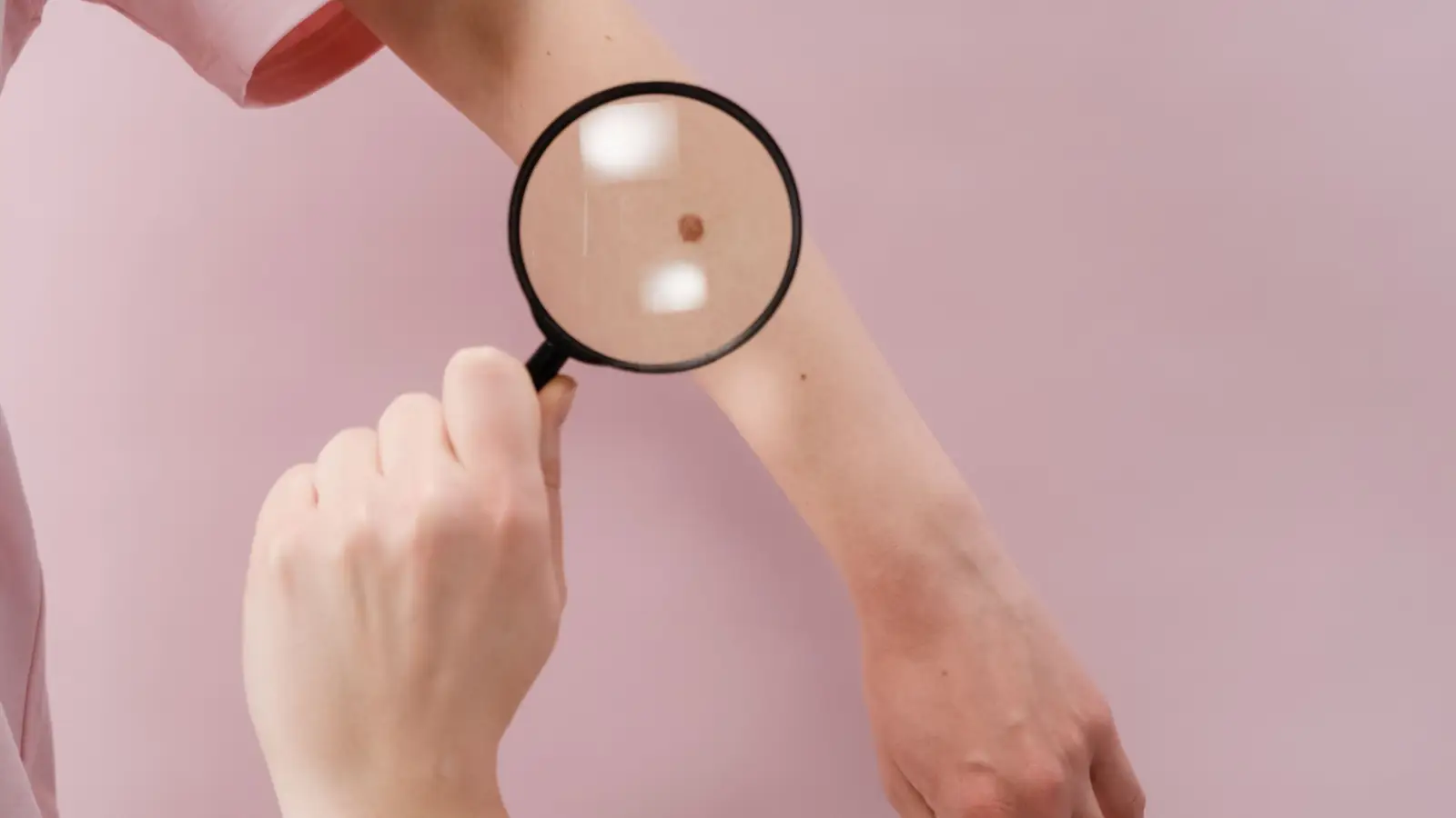
This is the narrow deep window: 755 nm energy concentrates its destruction in melanin-dense cells, leaving adjacent tissue essentially untouched — provided that the pigment forms a thin, well-demarcated target. When the window widens, as it does in melasma-prone or melanin-rich skin, that precision collapses and the heat spreads far enough to ignite an inflammatory response that darkens the very area the patient wanted lightened.
The 755 nm Peak and Selective Photothermolysis
The alexandrite laser’s wavelength sits near the melanin absorption maximum, so it can be calibrated to heat only structures that contain melanosomes. Selective photothermolysis is the mechanism: a pulse short enough — nanosecond or millisecond, depending on the handpiece — delivers energy faster than thermal relaxation can spread it beyond the target. Lentigines and freckles, which concentrate melanin in a shallow epidermal compartment, are destroyed while the collagen and keratinocytes around them stay cool.
That same absorption efficiency means every extra unit of melanin in the normal epidermis also captures some energy. In skin types with higher constitutive pigment, the baseline heating can approach a threshold where subclinical injury triggers melanogenesis, and that is the first crack in the window.
The Fitzpatrick Filter
Fitzpatrick I–III skin has a low to moderate starting melanin load. The contrast between a pigmented spot and the surrounding skin is large enough that the laser can treat the lesion without appreciably affecting the background. At type IV, background melanin begins to compete for energy, and the safety margin shrinks. Type V and VI skin have such high melanin density that the 755 nm pulse acts almost indiscriminately, delivering a thermal insult to the entire treatment area.
That is why alexandrite is rarely, if ever, used on darker skin for pigmented lesions. The risk of PIH and hypopigmentation is not a statistical outlier; it is a predictable outcome of the physics. Even a type IV patient may be treated only after a test spot and at conservative fluences, and some clinics decline outright. The filtering decision is binary at the extremes and tight in the middle.
Lentigines and Freckles: Where the Window Fits
Solar lentigines have dense, basal-layer melanin with occasional superficial dermal extension, but the lesion is thin and sharply bordered. Freckles are even more superficial, with a high density of melanin in a small area. Flat seborrheic keratoses, though not strictly pigmentation disorders, also respond because their melanin component can be ablated by the 755 nm pulse.
Under a Wood’s lamp or dermatoscope, these lesions enhance and remain crisp-edged. That crisp edge signals that the pigment is primarily epidermal and will react to a melanin-targeting wavelength. The clearance after two to three sessions is reliable; a single session often produces visible fading immediately.
Melasma: Why the Window Closes
Melasma is fundamentally different. Its melanin is not packed into a solitary nest; it is smeared through the epidermis and dermis, often with a vascular and hormonal driver. The 755 nm laser cannot distinguish melasma from normal skin because there is no sharp margin to exploit, and even a low fluence can heat enough melanocytes to provoke a rebound cascade. Melasma-prone skin is already in a state of hyperactivity, and thermal injury is frequently the trigger for a flare.
Consequently, a dermatologist who reaches for alexandrite when melasma is present is ignoring the biologic substrate. The laser may flatten the patch temporarily, but the rebound is usually worse. Topicals, oral tranexamic acid, and low-fluence 1064 nm lasers with fractionated delivery have a more rational basis for melasma. The narrow deep window that makes alexandrite elegant for a solar lentigo makes it a blunt instrument for melasma.
Post-Inflammatory Hyperpigmentation as the Deciding Risk
PIH is the darkening that follows any skin injury, and the threshold for that injury is far lower in skin of color. When a 755 nm pulse heats normal epidermal melanin, even if no blister forms, the melanocytes can interpret the event as damage and begin producing excess pigment that gets dumped into the dermis. What results is a stubborn, diffuse stain that can last for months and often appears worse than the original lesion.
A dermatologist evaluating candidacy will ask about any history of dark spots after acne, cuts, or previous procedures. A positive history in a Fitzpatrick III patient still allows treatment, but with extra caution and a mandatory test spot. In type IV, that history shifts the risk-benefit calculation enough that prudent clinicians often look for a different laser. In V and VI, the risk approaches near-certainty, and alexandrite is effectively contraindicated.
A recent tan, even in a type II patient, temporarily loads the epidermis with extra melanin and must be waited out. The skin must be at its baseline color, not its sun-exposed color, when the pulse fires.
The Consultation Sequence
Identify the Lesion Type
A dermatoscope or Wood’s lamp is the first tool. Epidermal pigment darkens under Wood’s light and stays well-defined; dermal or mixed pigment blurs or disappears. If the border fades under the lamp, the target is unlikely to respond cleanly to 755 nm and may be melasma or a deeper process.
Map the Skin Type and Tanning History
Beyond the Fitzpatrick number, the dermatologist asks: Do you burn or tan? Have you had laser or peel treatments before? Any reaction? How easily do you scar or develop dark marks from minor injuries? These questions refine the risk profile beyond a simple scale.
Review Medications and Topicals
Retinoids, isotretinoin, photosensitizing drugs, and recent chemical peels can amplify the laser’s effect and delay healing. The standard is to wait six months after oral isotretinoin and to pause topical retinoids several days before treatment. Any deviation from those timelines widens the risk.
Test Spot When Uncertainty Exists
If the Fitzpatrick type is borderline, the lesion looks slightly odd, or the patient reports a tendency toward PIH, a small test spot is treated and observed for two to four weeks. The absence of PIH or hypopigmentation in that spot is the only reliable indicator that a full treatment will be safe. No verbal reassurance replaces it.
Realistic Expectations
Even ideal candidates should expect two to three sessions spaced four to six weeks apart for lentigines. Freckles fade quickly but return with sun exposure; maintenance sessions once or twice a year are typical. Sunscreen is not a suggestion — it is the single most important determinant of whether the result holds.
The Treatment Experience and Aftercare
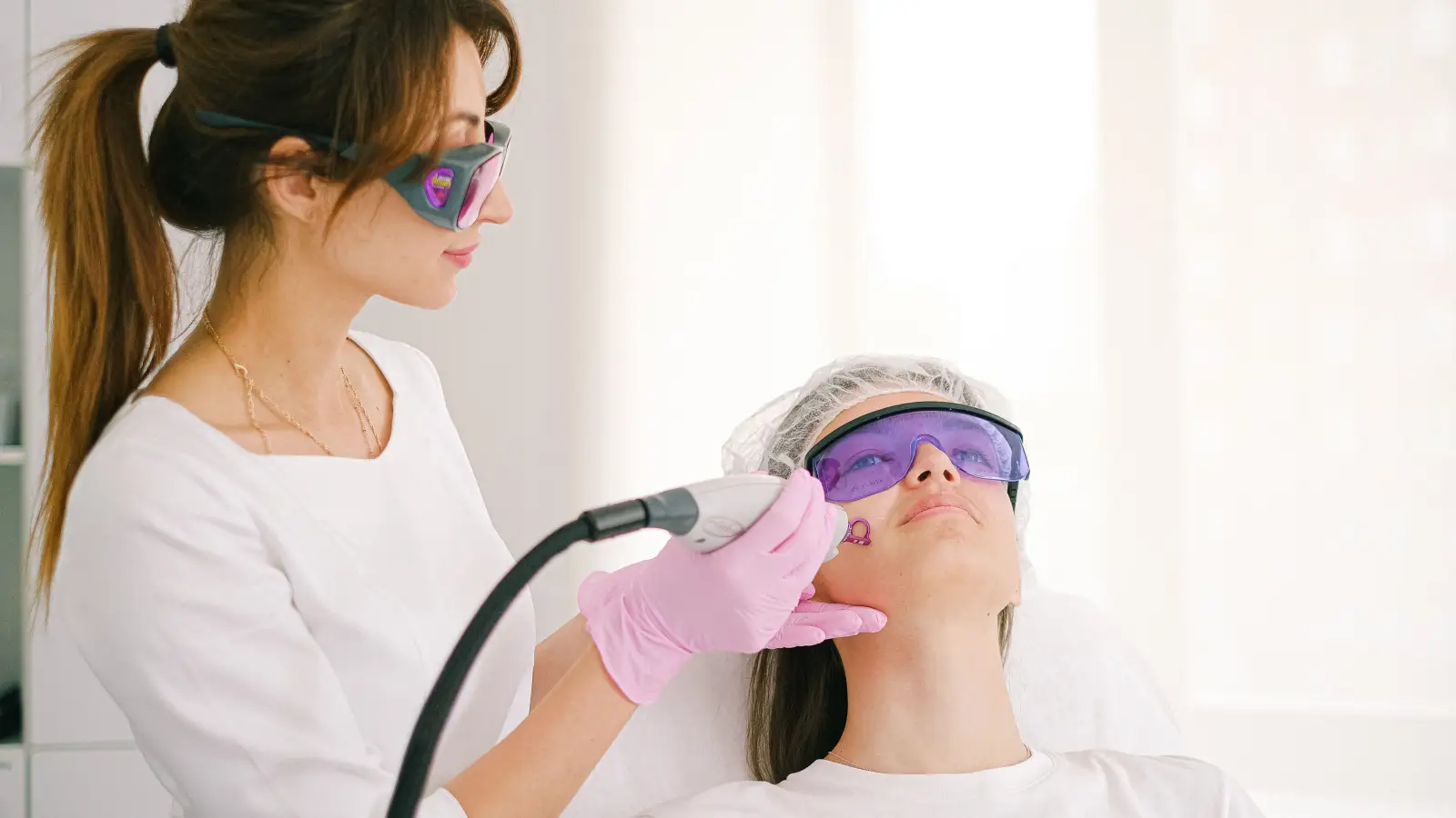
Each pulse feels like a snap and a brief heat, mitigated by a cooling tip. The treated spot turns white or ashy — a sign the pigment absorbed the energy — then darkens and crusts over the next few days. The crust flakes in one to two weeks, revealing lighter skin underneath.
Aftercare is uncomplicated but non-negotiable: a protective ointment to keep the area moist, avoidance of rubbing or picking, and strict sun protection. Broad-spectrum SPF 50+, wide-brimmed hats, and avoiding peak sun for four to six weeks post-treatment are baseline. Any sun exposure during the healing phase can repigment the area or trigger PIH, effectively zeroing out the benefit.
The Narrow-Deep-Window Summary
- Alexandrite’s 755 nm wavelength targets melanin with high selectivity, making it effective for discrete, superficial epidermal pigment in light skin.
- The dermatologist’s first decision is skin type: types I–III are candidates; type IV is borderline and demands a test spot; types V–VI are excluded.
- Lesion type is the second filter: solar lentigines, freckles, and flat seborrheic keratoses work; melasma and PIH do not.
- The risk of post-inflammatory hyperpigmentation drives almost every exclusion, especially in darker skin or when melanin is not confined.
- A Wood’s lamp, thorough history, and a test spot are the practical tools that confirm the narrow deep window is the right shape.
When the Window Holds — and When It Doesn’t
The alexandrite laser can clear a sunspot with a handful of short sessions and minimal downtime, but only when the skin and the lesion match its physical requirements. The single most useful step is an in-person assessment with a board-certified dermatologist who will examine the spots under magnification, determine Fitzpatrick type without guesswork, and rule out melasma. That examination — not a photo, not a phone consultation — is where the narrow deep window is measured. If the window fits, the result is predictable. If it doesn’t, pushing ahead anyway turns a cosmetic annoyance into a months-long pigmentation problem.
Frequently Asked Questions
Does alexandrite laser removal hurt?
Most people describe each pulse as a quick rubber band snap with heat. Cooling built into the device keeps it tolerable, and topical numbing is rarely needed.
How much does alexandrite laser treatment cost per session?
Cost varies by location and area size, but treating a few spots on the face or hands typically runs $150 to $400 per session in the U.S., with discounts for packages.
How is alexandrite different from a Q-switched Nd:YAG laser for pigmentation?
Nd:YAG at 1064 nm has much less melanin absorption, so it’s safer for darker skin and is often used for melasma and deeper pigment. Alexandrite’s 755 nm is faster for superficial spots on light skin but carries substantially more risk in skin of color.
Can alexandrite laser treat post-inflammatory hyperpigmentation itself?
No. PIH is often a diffuse pigment process that melanin-targeting lasers can worsen. First-line treatments are topicals like hydroquinone or azelaic acid, and low-fluence lasers only under careful supervision.
Is alexandrite laser safe on the face?
Yes, for the right candidate (types I–III with epidermal lentigines or freckles) the face is one of the most treated areas and heals well when sun protection is strict afterward.
What should I avoid before treatment?
Sun exposure, tanning beds, and self-tanners for four weeks; retinoids and exfoliating acids for several days; and any photosensitizing medications as directed by a physician. The skin must be at its natural color — not tanned — at the time of treatment.
Pigment Curious
We are people who want to better understand skin pigmentation, dark spots, and how different skin tones respond to skincare and professional treatments. We look beyond simple beauty claims, focusing on scientific evidence, clear explanations, and practical knowledge that helps people make more informed decisions about their skin.