Why Picosecond Alexandrite Isn't Always Better for Spots
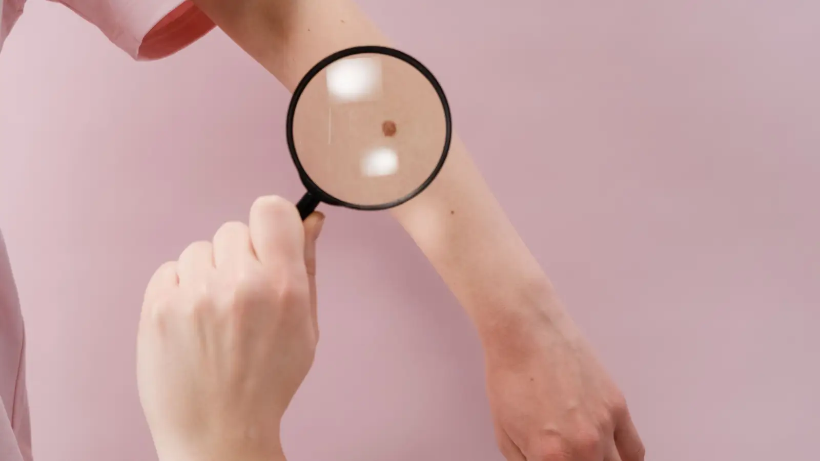
The logic seems sound: a 750-picosecond pulse releases energy so rapidly that melanosomes shatter before heat can spread, leaving surrounding tissue untouched. It’s the reason many patients—and even some clinicians—assume that picosecond alexandrite is an upgrade for every dark spot. But that assumption breaks down when you account for lesion geography. A dense lentigo isn’t a uniform layer of individual melanosomes; it’s a three-dimensional pigmented scaffold that can span several millimeters. A pulse confined to micron-scale disruption may not cover the entire volume. For spots that are large, thick, and darkly pigmented, a Q-switched nanosecond alexandrite at the same wavelength often achieves better clearance in fewer sessions—not because it’s more powerful, but because its shockwave is physically wider.
The idea that a shorter pulse always improves clearance is a classic example of mistaking a single variable for a full mechanism. What matters is not just fragmentation fineness but whether the disruptive field envelops the whole lesion. This article dissects the photomechanical differences, builds a spot-morphology matrix, and shows why skipping the pico in favor of the nano may be the smarter clinical choice for dense lentigines.
The Case Against Pico for Large Dense Spots
For a well-circumscribed solar lentigo measuring 6 mm or more—dark, slightly raised, and heavily pigmented—a Q-switched nanosecond alexandrite (755 nm, 5–20 ns) delivers a higher probability of complete clearance after one or two sessions. Picosecond devices frequently undertreat these lesions: they fragment individual melanosomes beautifully but leave the larger pigment infrastructure intact, resulting in persistent brown cores that require multiple additional visits. If the spot is fine, recalcitrant, and faint, picosecond may find its niche; for every other common presentation, nanosecond should be the default starting point.
A Spot-Morphology Lens: Picosecond vs Nanosecond Alexandrite
| Picosecond Alexandrite | Q-Switched Nanosecond Alexandrite | |
|---|---|---|
| Pulse duration | 450–750 ps | 5–20 ns |
| Dominant photomechanical effect | Stress-confined, sub-micron fragmentation | Thermal cavitation with broader acoustic disruption |
| Approximate disruption zone per absorption site | A few microns; gap-bridging is limited | Tens of microns; covers adjacent clusters |
| Spot types it handles best | Fine, stippled lentigines <5 mm; residual pigment after other modes | Dense, well-demarcated lentigines >5 mm; thick epidermal pigment |
| Typical sessions to clear a large lentigo | 3–5+ (frequent need for touch-ups) | 1–3 (higher single-session clearance) |
| Thermal spread | Negligible; essentially athermal breakup | Moderate; can be managed with fluence and spot size |
| PIH risk in Fitzpatrick IV–VI | Lower when fluence is kept low | Higher if aggressive settings used; requires test spots |
| Approx. U.S. cost per session | $250–$500 | $150–$400 |
How the Two Pulse Regimes Actually Break Pigment
Picosecond Alexandrite: Localized Disruption That May Skip the Scaffold
Picosecond 755 nm pulses are shorter than the stress relaxation time of a melanosome. That condition (stress confinement) creates an intense, brief pressure wave that fractures melanin into nanoparticles. The advantage is clear: almost no heat diffuses into adjacent keratinocytes, which appeals strongly for treating pigment in skin of color where thermal injury can trigger PIH.
Yet the fragmentation zone is extremely narrow. In a dense lentigo, melanosomes aggregate into large clusters held together by cellular membranes, melanophages, and thickened stratum corneum. A picosecond shockwave will crack individual melanosomes but often fails to propagate across cluster boundaries. Clinically, this manifests as a spot that lightens unevenly—a central island of pigment remains. Multiple sessions can gradually nibble away at the residual pigment, but the efficiency drops when compared to a treatment that can demolish the whole cluster at once.
Q-Switched Nanosecond Alexandrite: A Broader Blast That Covers the Lesion
Nanosecond pulses at the same 755 nm dwell in the intermediate regime where pulse duration exceeds the acoustic relaxation time but remains short enough to avoid bulk tissue heating. Here, the mechanism shifts toward rapid thermal vaporization accompanied by cavitation bubble formation. The expanding and collapsing bubble produces a mechanical disruption radius that is an order of magnitude larger than the picosecond case—tens of microns rather than a few.
That broader zone is why a single nanosecond session can turn a 7-mm lentigo into an immediate frosting endpoint that heals clear. The tradeoff: the thermal footprint is larger, and in darker skin types, that heat can stimulate melanocytes, potentially causing PIH. The answer isn’t to abandon nanosecond, but to apply it with deliberate parameter selection—larger spot sizes, lower fluences, and careful test spots. When those precautions are observed, nanosecond often clears dense lentigines where picosecond would have required serial re-treatment.
Three Criteria That Actually Dictate Clearance
Fragmentation Zone vs Lesion Diameter
Clearance success depends on whether the mechanical disturbance field encompasses the entire lesion thickness and width. A picosecond pulse’s zone of a few microns is adequate for fine dust-like pigmentation but undersized for a lentigo where melanosome clusters can be 20–50 µm across. Nanosecond pulses cover that scale, which explains why many practitioners observe that dense spots clear faster with a Q-switch despite the older technology.
Thermal Modulation of Melanocytes
Heat isn’t universally harmful. Mild thermal injury to hyperactive melanocytes at the base of a lentigo may reduce recurrence—a subtle benefit of nanosecond’s moderate heating. Picosecond avoids that heat, which is excellent for melasma but may leave the melanocyte population intact and ready to re-pigment, especially in lentigines driven by cumulative sun exposure. The matrix must consider not only instant clearance but long-term durability.
Cumulative Time and Cost
A typical large lentigo resolved in two nanosecond sessions at $200 each costs $400 total; a similar spot requiring five picosecond sessions at $300 each racks up $1,500 plus months of visits. The additional sessions also prolong the risk of post-inflammatory changes and patient attrition. When the metric is “sessions to satisfactory clearance for a dense spot,” nanosecond often wins, a point that gets lost in the marketing noise around pulse duration.
Choosing Treatment by Spot Morphology
Predominant feature: dark, thick, well-defined lentigo >5 mm
Begin with Q-switched nanosecond alexandrite, 755 nm, starting at 2.5–4 J/cm² with a 3–5 mm spot. Aim for immediate frosting. A single pass frequently clears the lesion entirely; a second session at 4–6 weeks handles any residual fleck.
Predominant feature: fine, stippled, or stubborn residual lentigo <5 mm
Picosecond alexandrite offers an advantage here. Use low fluence and multiple passes to lift light pigment without generating a thermal footprint. This morphology is where the ultra-short pulse justifies its premium.
Predominant feature: multiple lentigines on deeply pigmented skin
Start with a conservative approach. Low-fluence picosecond or careful long-pulse nanosecond can work, but test spots are essential. The guiding principle: on Fitzpatrick IV–VI skin, undertreatment with a safe modality is always preferable to aggressive treatment that triggers PIH. In many such cases, picosecond’s minimal heat profile gives it a safety edge even if clearance may be slower.
Questions That Come Up Around Pulse Duration
Does picosecond alexandrite hurt less?
Sensation is similar when topical anesthetic is used; both create a sharp snap. Some patients perceive picosecond as a faster “snap” and nanosecond as a deeper, lingering heat. Pain is rarely the pivotal factor in choosing one over the other.
Can picosecond prevent all PIH?
No device eliminates PIH risk in darker phototypes. Picosecond reduces conduction heat, which lowers the probability, but cavitation alone can still disrupt melanocytes enough to trigger inflammation. Post-treatment cooling, strict sun avoidance, and melanin-suppressive topicals remain non-negotiable.
Is it possible to combine nanosecond and picosecond in one session?
Certainly. Clinicians working on mixed spot typography may use nanosecond for the dense core and picosecond for the lighter periphery or adjacent lentigines. The choice is guided by real-time lesion endpoint and the provider’s experience reading skin response.
The Physics Doesn’t Care About Marketing
Shorter pulses are not an unqualified upgrade. For the most common concern—a dark, thick solar lentigo large enough to be embarrassing—the broader fragmentation zone of a Q-switched nanosecond alexandrite delivers faster, more complete clearance than the ultra-localized pop of a picosecond pulse. Before committing to a series of pico treatments, examine the morphology: if the spot is dense and well-demarcated, the older technology may be the better tool.
Pigment Curious
We are people who want to better understand skin pigmentation, dark spots, and how different skin tones respond to skincare and professional treatments. We look beyond simple beauty claims, focusing on scientific evidence, clear explanations, and practical knowledge that helps people make more informed decisions about their skin.