What Dermatologists Check About Your Skin Tone Before Treating Spots
Dermatologists check your Fitzpatrick skin type before touching a spot because that single number predicts whether you’ll heal clean or trade one mark for a bigger, darker one. It is not a formality. On Fitzpatrick I, a brown spot is often a sun-induced solar lentigo that responds briskly to cryotherapy or a 532 nm laser. On Fitzpatrick V, a spot that looks identical to the naked eye is frequently melasma—deeper, hormonally driven, and primed to flare into post-inflammatory hyperpigmentation (PIH) if hit with the wrong energy. The same lesion, different skin tones, opposite treatment paths. That is the core logic a dermatologist applies before anything touches your face.
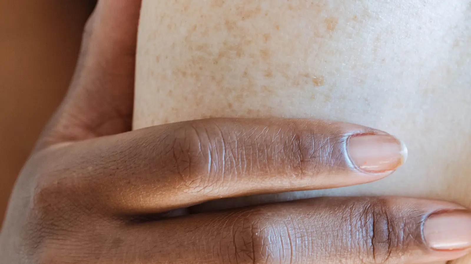
The Fitzpatrick scale maps how melanin behaves in your skin: how much you have, where it sits, and how reactive your melanocytes are when inflamed. This data point locks in the safety margin for lasers, chemical peels, and even topical actives. When a protocol respects that margin, a spot fades. When it ignores tone, the result is often worse than the original complaint. Now the step-by-step framework.
Why Spot Diagnosis and Fitzpatrick Type Are a Single Decision
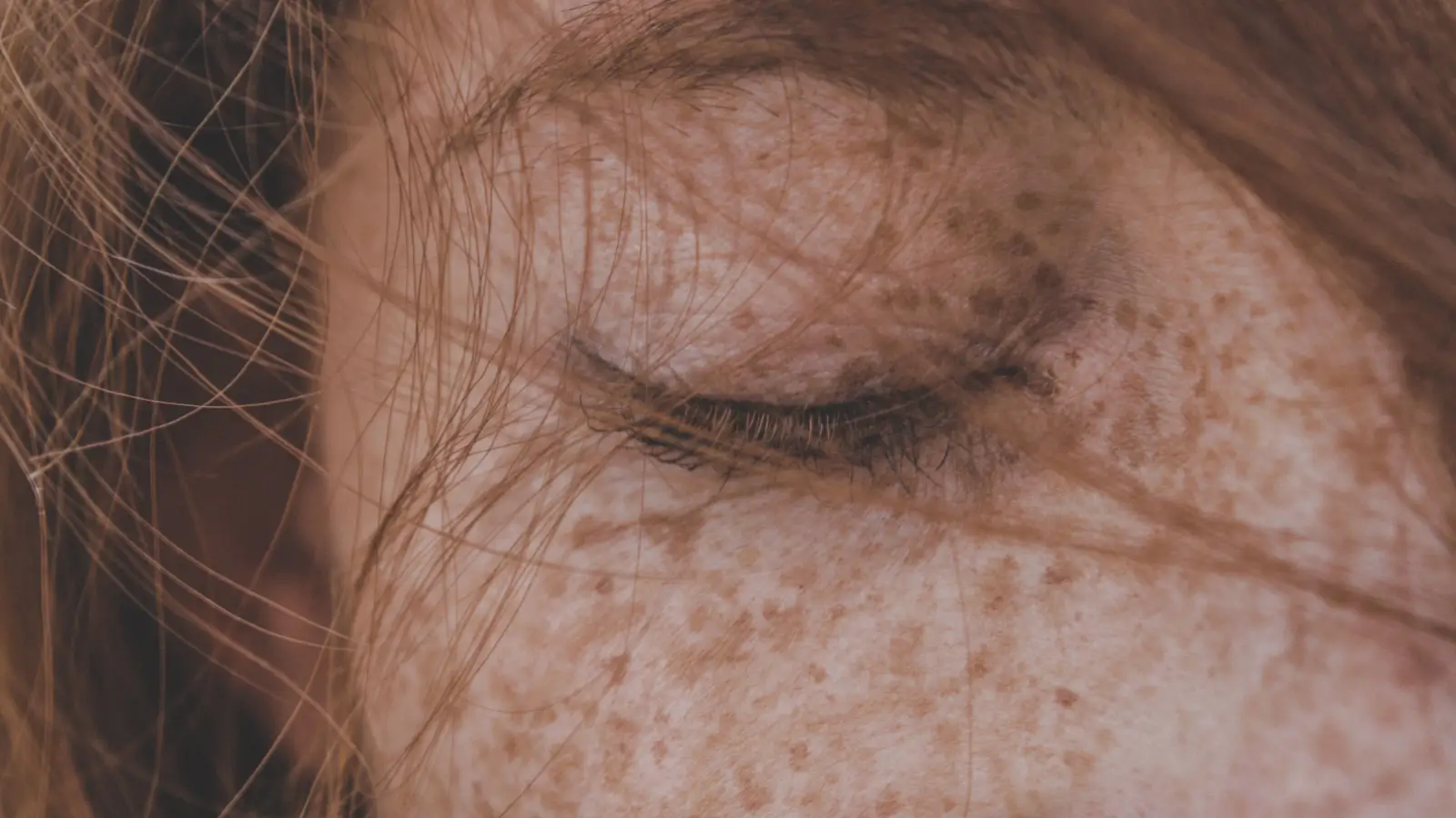
A flat brown patch on the cheek is not one diagnosis. It could be a solar lentigo (superficial, UV-driven), melasma (deep, hormonal), or PIH from an old breakout. On Fitzpatrick I–II skin, a solar lentigo shows up as a crisp, light-brown mark that responds predictably to IPL or cryotherapy. On Fitzpatrick IV–VI skin, that same macular area often turns out to be melasma—diffuse borders, pigment sitting in the dermis, and completely incompatible with IPL. Treating the wrong spot identity on the wrong skin tone creates a cascade: hyperpigmentation, textural changes, and sometimes hypopigmented scars.
This is not theory. A Wood’s lamp or dermatoscope reveals where pigment actually lives—epidermis or dermis—and that answer, cross-referenced with your Fitzpatrick category, dictates whether a laser is even an option. Spot diagnosis and tone classification are a single decision. You cannot correctly treat one without the other.
The Three-Step Skin Tone Safety Check
Dermatologists follow a linear sequence that prevents the most expensive mistakes: burns, scars, depigmentation, and months of PIH. Each step locks in a variable the next step cannot correct if skipped.
Step 1: Diagnose the Spot Using Tone-Aware Visual Cues
The same biological lesion looks different across the Fitzpatrick spectrum. Melasma on Fitzpatrick II can appear as a faint grey-brown veil. On Fitzpatrick VI, it commonly presents as deep brown to blue-grey patches because dermal melanin scatters light differently. A dermatologist reads the lesion’s borders, color uniformity, Wood’s lamp fluorescence, and epidermal versus dermal placement—all through the filter of your underlying pigmentation.
Done well, this step outputs a working diagnosis and a map of pigment depth. Without it, you are treating a picture, not the pathology.
Step 2: Classify Fitzpatrick Type With Absolute Precision
Fitzpatrick typing is not a guess. The dermatologist asks about your sunburn history and examines skin that has not seen UV—typically the inner arm. The scale runs from I (always burns, never tans) to VI (deeply pigmented, never burns). This number is a proxy for melanocyte reactivity and melanin density, the two variables that determine injury threshold.
This step comes second deliberately: the spot diagnosis is already known, so the dermatologist now sets the boundary for how much thermal or chemical stress the skin can absorb without triggering PIH. A Fitzpatrick I patient with a solar lentigo can safely take a high-fluence Q-switched laser or a medium-depth TCA peel. A Fitzpatrick IV patient with the same spot diagnosis must switch to a longer-wavelength laser, conservative fluence, and often a pre-treatment with a tyrosinase inhibitor.
Done well, this step turns a subjective skin tone into an objective, treatment-guiding number.
Step 3: Match the Treatment to the Skin’s Pigment Risk Profile
With the spot identity and Fitzpatrick number fixed, the dermatologist selects the exact modality and parameters that fit the tone’s safety ladder:
- Fitzpatrick I–II: Broad treatment window. IPL, cryotherapy, deep peels, and short-wavelength lasers are viable. Primary risk is hypopigmentation from over-treatment, not PIH.
- Fitzpatrick III–IV: Melanin content rises. Lasers must shift to longer wavelengths (1064 nm Nd:YAG, not 755 nm alexandrite), longer pulse durations, and epidermal cooling. Medium-depth peels are usable but at lower concentrations and with cautious application time.
- Fitzpatrick V–VI: The margin for error is razor-thin. Topical depigmenting agents are first-line. If lasers are used, they are low-fluence, large-spot, long-pulse Nd:YAG with aggressive contact cooling, or non-ablative fractional at conservative densities. A test spot in a concealed area is mandatory—treating the full face without it is negligence.
Done well, the treatment slots into a narrow band where it clears the target pigment without waking the surrounding melanocytes.
The Framework in Motion: One Spot, Two Skin Tones, Two Outcomes
A flat, dime-sized brown patch sits on the cheek. Here is how the same spot appearance triggers two completely different paths.
Fitzpatrick II path: Dermoscopy shows a sharply defined, light-brown lesion limited to the epidermis. Wood’s lamp confirms no dermal extension. History: decades of sun exposure, no hormonal triggers. Diagnosis: solar lentigo. Fitzpatrick type II means minimal PIH risk. The dermatologist selects a single cryotherapy session or a 532 nm Q-switched laser. The spot clears in weeks.
Fitzpatrick V path: The same brown patch is actually indistinct at the edges. Under Wood’s lamp, pigment forms a net-like pattern reaching the dermis. History: appeared during pregnancy, worsens with heat. Diagnosis: dermal melasma. A cryotherapy wand or IPL would burn the skin and ignite PIH that could last a year. The dermatologist instead prescribes a triple-combination cream and only schedules low-fluence 1064 nm Nd:YAG after a two-week test spot proves the skin can handle it. The framework turned an impending disaster into a controlled plan.
Where the Framework Fails in Practice
Treating Before the Spot Diagnosis Is Firm
Patients often self-label a spot as “sun damage” and walk in requesting a laser. If the provider treats without a Wood’s lamp exam or dermatoscopy, the lesion could be anything—seborrheic keratosis, nevus, or lentigo maligna. Add a dark skin tone to that scenario, and the failure compounds: an aggressive laser on undiagnosed lentigo maligna delays cancer detection, and the ensuing PIH buries the clinical signs. Step one is non-negotiable.
Skipping the Fitzpatrick Ladder When a Laser Seems “Mild”
Not all lasers are equal, and no laser is safe just because a medspa markets it as “for all skin types.” A 755 nm alexandrite laser that feels gentle on Fitzpatrick II can vaporize melanin-rich keratinocytes on Fitzpatrick IV, leaving atrophic scars or leopard-spot hypopigmentation. The right wavelength, pulse duration, and fluence change with every rung of the Fitzpatrick scale. Applying a one-size-fits-all protocol is how healthy skin gets traded for permanent damage.
Overlooking the Aftermath: PIH Is the Real Treatment Failure
Even a correctly chosen laser or peel can fail if post-procedure care ignores tone-specific needs. Fitzpatrick IV–VI skin requires strict, broad-spectrum mineral sunscreen use, cessation of physical exfoliation for weeks, and often a pre- and post-treatment tyrosinase inhibitor. When a provider omits that instruction, inflammation from the treatment itself triggers new melanin production. The original spot fades, but a darker shadow takes its place—and that outcome was entirely preventable if the framework had been carried through to the healing phase.
Quick Reference: Skin Tone-Safe Spot Treatment
| Evaluation Step | Goal | Key Action |
|---|---|---|
| Spot Identification | Distinguish solar lentigo, melasma, PIH, or other | Wood’s lamp or dermoscopy; map border pattern and pigment depth relative to skin background |
| Fitzpatrick Classification | Quantify melanin content and injury response | Burn/tan history plus non-sun-exposed skin assessment |
| Treatment Matching | Select modality within that tone’s safety profile | Align wavelength/fluent, peel depth, or topical protocol with Fitzpatrick-specific risk |
FAQ
Can I treat spots at home without knowing my Fitzpatrick type?
Over-the-counter fading creams containing azelaic acid, kojic acid, or low-percentage vitamin C can be safe across the spectrum, but PIH risk rises if you add high-strength exfoliating acids. Without a Fitzpatrick classification, you lack the guardrail that tells you where the irritation threshold sits. A dermatologist typically types your skin in minutes; that single data point prevents weeks of trial-and-error.
Is the Fitzpatrick scale the only thing that matters for laser safety?
No. Spot depth, recent tanning, and ethnic background (which influences melanocyte lability) also matter. The Fitzpatrick scale is the starting reference, not the complete decision tree.
Can a dermatologist safely treat spots on very dark skin?
Yes, with precise protocols. Low-fluence 1064 nm Nd:YAG lasers, non-ablative fractional resurfacing at conservative densities, and topical lighteners are standard tools. Success requires a test spot, aggressive cooling, and understanding that zero risk is impossible—but managed risk is achievable.
What happens if a chemical peel is too strong for my skin tone?
A peel that reaches too deep in medium-to-dark skin can cause post-inflammatory hyperpigmentation larger and darker than the original spot. In some cases, it destroys melanocytes altogether, leaving permanent hypopigmented patches. That is why dermatologists start with superficial peels at low concentration and only increase strength after seeing the healing response.
Start With the Spot Diagnosis—Your Skin Tone Defines the Path
The three-step safety check—diagnose the spot, classify the Fitzpatrick type, match the treatment—is the only sequence that keeps spot removal from becoming spot creation. If you act on one point today, it is this: before any laser, peel, or potent topical, have a dermatologist identify both the lesion and your Fitzpatrick type side-by-side. A correct spot treatment is worthless if it ignores the skin it’s being applied to.
Pigment Curious
We are people who want to better understand skin pigmentation, dark spots, and how different skin tones respond to skincare and professional treatments. We look beyond simple beauty claims, focusing on scientific evidence, clear explanations, and practical knowledge that helps people make more informed decisions about their skin.