Your ‘Melasma’ Might Actually Be Post-Inflammatory Hyperpigmentation
If you’ve been told you have melasma but the dark patches do not respond to treatment—or they flare after a peel or laser—post-inflammatory hyperpigmentation (PIH) may be the real diagnosis. These two conditions look similar at a glance, but they form through entirely different mechanisms. That difference matters because the standard first-line approach for one can worsen the other.

Melasma is a hormonally driven pigmentary disorder, activated by estrogen, progesterone, and UV or heat exposure. PIH is a local wound-healing response: any inflammation, from a pimple to a laser burn, can cause melanocytes to overproduce pigment at the site of injury. Their distribution makes the distinction clearer: melasma presents as symmetrical, blotchy patches across the cheeks, forehead, and upper lip; PIH follows the exact footprint of a past lesion. And they respond unevenly to key actives. Hydroquinone suppresses pigment in melasma but can irritate vulnerable PIH skin and darken the spot. Tranexamic acid addresses the vascular component of melasma and has no established role for uncomplicated PIH. Azelaic acid treats both safely because it combines anti-inflammatory and mild tyrosinase-inhibiting effects. Recognizing which condition is present prevents a treatment from becoming the trigger for more discoloration.
Which One Is It? A Situational Breakdown
| Scenario | Likely Diagnosis | Reason |
|---|---|---|
| Symmetrical brown patches appear on both cheeks, forehead, or upper lip, often after hormonal changes (pregnancy, oral contraceptives) | Melasma | Hormonal influence and symmetrical pattern are classic melasma markers |
| A dark mark sits exactly where a pimple, cut, or chemical peel burn was located | PIH | Site-specific discoloration tied to prior inflammation |
| Pigment worsens after a strong peel or laser meant to fade it | PIH or iatrogenic melasma flare | Procedure-induced inflammation can spark new PIH or aggravate existing melasma |
| The discoloration fades noticeably in winter and darkens in summer | Both, but especially melasma | UV and heat are major melasma triggers; PIH also darkens with sun but usually correlates more with injury |
When Symmetrical Patches Cover the Cheeks and Forehead
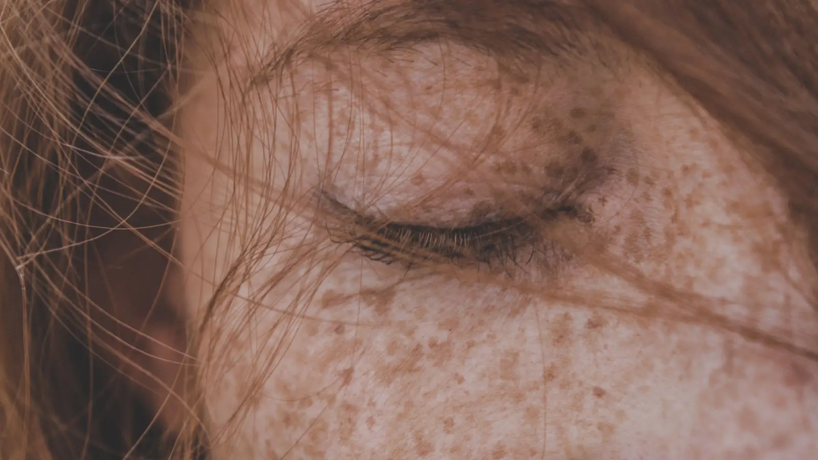
This is the textbook presentation of melasma. The symmetrical, butterfly-like distribution on sun-exposed skin—often seen in centrofacial, malar, or mandibular patterns—is a direct consequence of hormonal sensitivity. Estrogen and progesterone stimulate melanocytes, and UV radiation plus visible light multiply that effect. In melasma, pigment deposits are found in both the epidermis and dermis, often with a faint vascular redness beneath, which is why tranexamic acid (which reduces vascular leakage and melanocyte-stimulating factors) can be a targeted part of treatment.
The medication logic follows two tracks: blocking tyrosinase and quieting the vascular input. Hydroquinone 4% remains the most potent topical lightener, typically compounded with tretinoin and a corticosteroid to limit irritation. Oral tranexamic acid at 250 mg twice daily or topical formulations reduce relapse by addressing dermal vasculature. Azelaic acid 15–20% is the primary alternative during pregnancy, when hydroquinone is off-limits. Sun protection must be the strictest part of the routine: tinted mineral sunscreens with iron oxides block visible light as well as UV, and heat exposure should be minimized because it can re-trigger the pathway on its own.
Choosing to manage this as melasma means accepting that the condition is chronic and will likely recur. What you give up is the option to use deep peels or ablative lasers for faster clearance—those procedures often provoke rebound pigment, especially on melanin-rich skin, making them a poor trade for melasma.
When a Spot Lingers After a Pimple or Skin Injury
This marks the classic story of post-inflammatory hyperpigmentation. Any inflammatory event—acne, an ingrown hair, a scratch, a poorly tolerated product, even a sub-ablative laser—can leave a flat, brown or grayish mark directly over the area that healed. There is no hormonal driver, no symmetry, no tendency to spread. In darker skin tones, where melanocytes are inherently more reactive, a minor blemish can persist for months.
The treatment sequence is different. The first goal is to eliminate ongoing inflammation and maintain barrier integrity, not to force pigment suppression. Azelaic acid fits this need: it calms inflammation while inhibiting tyrosinase mildly, and it’s compatible with acne-prone skin where new lesions mean new PIH. Hydroquinone can be added once the skin surface is intact, but introducing it too early on disrupted skin often causes irritation that darkens the spot further. Tranexamic acid lacks a clear role because the vascular pathway it targets is not activated in simple injury-induced PIH. Topical retinoids, vitamin C, and gentle exfoliants (mandelic acid, low-concentration glycolic acid) speed cell turnover, but they must be introduced one at a time and at low frequency to avoid triggering a new round of inflammation.
Managing this as PIH yields a cause-specific plan that protects the skin’s healing process rather than overriding it. What you give up is any benefit from hormonal modulation or vascular-targeted agents, because those axes are not involved.
Where Melasma and PIH Overlap
There are several similarities that do not help in distinguishing the two, and you can stop weighing them as diagnostic clues.
Both conditions are forms of hyperpigmentation driven by excess melanin; both worsen with unprotected sun exposure, so high-SPF, broad-spectrum sunscreen is mandatory regardless of diagnosis. Both respond partially to tyrosinase inhibitors like kojic acid, arbutin, and azelaic acid, so a product containing these won’t point to one condition over the other. Both look darker on medium to deep skin tones because of higher baseline melanin. And both can take months to fade, particularly when dermal pigment is present. These parallels do not erase the fundamental differences in origin and treatment strategy, and they should not distract from the need for an accurate diagnosis before selecting a protocol.
Signs That Point Clearly to One Diagnosis
Some features act as a diagnostic switch; their presence practically settles the question.
Symmetrical, blotchy pattern on sun-exposed areas that worsens with heat and hormones — this is melasma. If the patches first appeared or intensified during pregnancy or after starting an oral contraceptive, and they spread symmetrically across the central face, cheekbones, or jawline, melasma is overwhelmingly likely. Dermoscopy typically shows a mixed epidermal-dermal pigment network with visible telangiectasias.
A dark mark or cluster that closely follows the outline of a healed lesion — this is PIH. When the timeline is clear (the spot formed where a pimple used to be, or exactly over a microneedling site) and the other side of the face shows no matching mark, the diagnosis is not melasma. Dermoscopy usually finds epidermal hyperpigmentation with a preserved pigment network and no vascular component.
Treating what looks like melasma with aggressive modalities—deep peels, high-fluence lasers—without confirming the diagnosis can transform a superficial PIH problem into a deeper, harder-to-treat pigment disorder. This risk is especially high on Fitzpatrick skin types IV–VI, where the threshold for procedure-induced PIH is low.
Frequently Asked Questions
Can a dermatologist distinguish melasma from PIH just by looking?
Usually, yes. A dermatoscope or Wood’s lamp reveals patterns—such as the reticular pigmentation typical of melasma or perifollicular sparing in some PIH—that are difficult to see with the naked eye. These tools often resolve an ambiguous history.
I used a melasma cream and the spots got darker. What went wrong?
If the real diagnosis was PIH, applying a strong active like high-concentration hydroquinone or tretinoin to post-inflammatory skin likely triggered irritation, which stimulated more melanin production. This is a common cycle of misdiagnosis.
Is laser safe for both melasma and PIH?
Safety depends on laser type, parameters, and skin tone. For melasma, even low-fluence Q-switched lasers can cause rebound hyperpigmentation; many dermatologists avoid lasers altogether on darker skin. For PIH, certain gentle devices (low-fluence 1927 nm thulium, picosecond lasers) may help, but only after inflammation has resolved, and the risk remains significant in melanin-rich skin.
Can I use tranexamic acid for post-inflammatory hyperpigmentation?
Tranexamic acid works by reducing plasmin activity and vascular endothelial growth factor, pathways relevant to melasma’s vascular component. Typical PIH from a blemish or cut does not activate that pathway, so tranexamic acid cannot be expected to help and is not part of standard PIH protocols.
Do I really need sunscreen indoors if I have melasma?
Yes. UVA and visible light pass through windows, and melasma is extremely sensitive to both. Tinted mineral sunscreens with iron oxides block visible light more effectively than untinted formulas, providing better protection indoor as well.
The most frequent misdiagnosis scenario—interpreting dark spots left over from old acne or a peel reaction as melasma—points to PIH as the actual problem when treatments fail. Before naming the condition, ask: Did these spots develop precisely where my skin was previously inflamed?
Pigment Curious
We are people who want to better understand skin pigmentation, dark spots, and how different skin tones respond to skincare and professional treatments. We look beyond simple beauty claims, focusing on scientific evidence, clear explanations, and practical knowledge that helps people make more informed decisions about their skin.