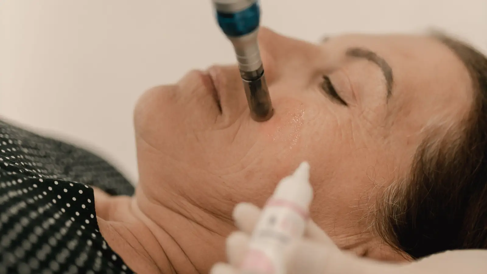
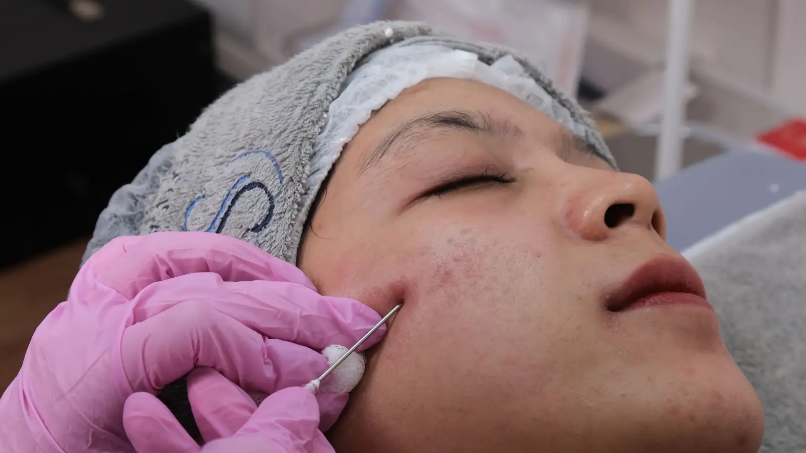
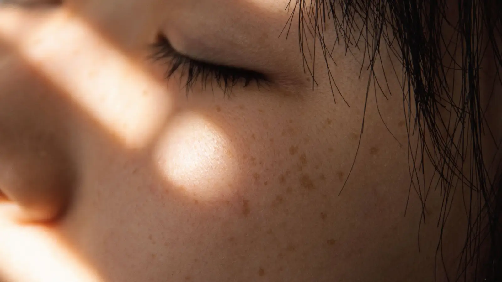
Microneedling for dark spots? Not all types respond.
Microneedling can lighten dark spots, but only when the pigment is epidermal. Dermal hyperpigmentation, especially on melanin-rich skin, often darkens after needling. The decision turns on two variables: where the pigment sits and how the skin’s melanocytes react to injury.
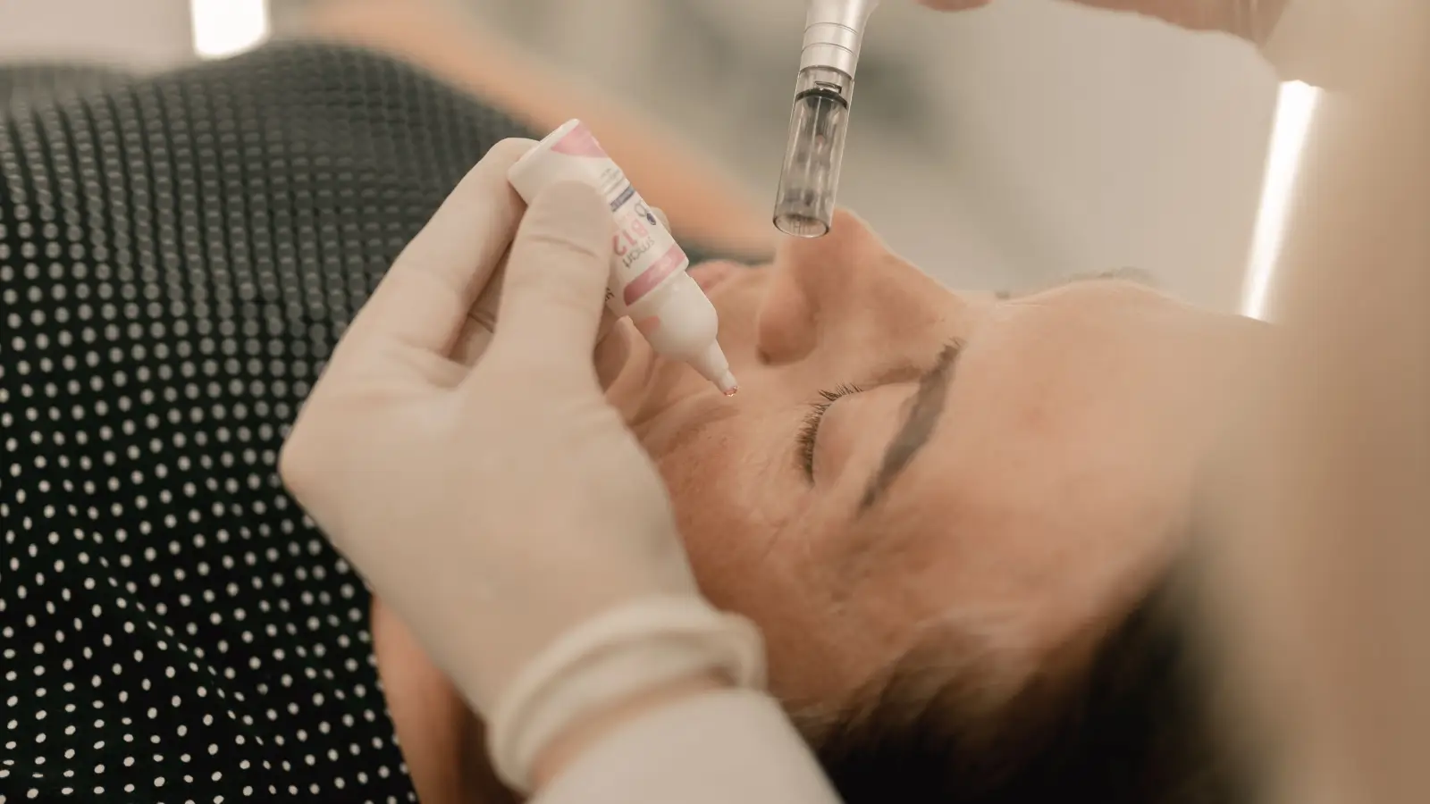
Intermediate practitioners hit a wall here because they’ve been taught that microneedling is “good for hyperpigmentation.” That overgeneralization collapses a dozen distinct pigment disorders into one. The Fitzpatrick scale and a pigment-depth classification are what break the logjam. On a Fitzpatrick V patient, a 1.0 mm needle over dermal melasma is not a treatment—it’s a trigger for stubborn, trackmark-shaped PIH.
Why Treating All Brown Spots as Interchangeable Fails
It seems reasonable to group every dark patch under one umbrella. Microneedling mechanically shatters melanin clumps and speeds epidermal turnover, so logic implies it should improve any discoloration. The false comfort here comes from ignoring etiology: solar lentigines, post-inflammatory hyperpigmentation, and melasma behave differently under identical mechanical stress.
The concrete damage occurs when needles cross into the dermis in conditions where melanocytes are already unstable. In dermal melasma, melanin sits inside macrophages deep in the dermis. Needling that layer provokes an inflammatory response that can intensify pigment production far beyond baseline. On Fitzpatrick IV–VI skin, this threshold is lower—the melanocyte’s alarm system fires with less provocation.
Experts replace the blanket approach with a pre-treatment classification. A Wood’s lamp or dermatoscope distinguishes epidermal (accentuated) from dermal (no accentuation) pigment. If the lesion is dermal, the first-line intervention is topical tyrosinase inhibition, and microneedling—if used at all—is restricted to 0.25 mm depth solely to drive those topicals into the skin. Needling is not deployed as a pigment-breaker in this scenario.
The Problem with Ignoring Fitzpatrick When Selecting Needle Depth
Protocols often set depth by treatment goal: 0.5 mm for rejuvenation, 1.0 mm for scars. When pigment clearance is the aim, the intermediate instinct is to go deeper—under the assumption that more mechanical disruption equates to more melanin removed. That assumption holds for epidermal pigment on low-Fitzpatrick skin, but it backfires once the dermis is involved or the melanocyte is easily provoked.
The damage appears as iatrogenic hyperpigmentation. Longer needles inflict microtrauma that melanin-rich skin interprets as a threat, laying down fresh pigment in the pattern of the punctures. Published clinical guidance on microneedling in Fitzpatrick IV–VI notes that PIH risk climbs sharply when needle length exceeds 1.0 mm without prior melanin suppression.
The expert move inverts the logic: let the pigment’s anatomical depth dictate needle choice. An epidermal lentigo on a Fitzpatrick II receives 0.25–0.5 mm. A suspected dermal melasma on Fitzpatrick IV never sees a needle longer than 0.25 mm—and only after weeks of topical pre-treatment. Depth is selected to barely reach the target, not to exceed it.
What Consistency Actually Requires: A Fixed Pre-Treatment and Depth Protocol
A 4–6 week evening course of a tyrosinase inhibitor (4% hydroquinone or 15% azelaic acid) for any Fitzpatrick III and above person before the first session. This suppresses baseline melanocyte activity and narrows the window for PIH.
Wood’s lamp mapping at every initial consult. Each patch gets labeled epidermal, dermal, or mixed; that label directly gates the treatment plan. A dermal classification removes standalone microneedling from the options.
Standard depth ceilings. The clinic defaults to 0.25 mm for pure epidermal lesions on any skin tone, permits 0.5 mm only when a mixed lesion shows strong epidermal dominance, and caps all hyperpigmented skin at 1.0 mm—even when the patient’s primary concern is acne scarring.
Patch testing on a small, hidden area with a 2–4 week observation window before committing to full-face treatment on any skin that has never been needled. The response at week two and week four decides whether to proceed.
How an Expert Decides When the Depth Is Unclear
If a Wood’s lamp cannot cleanly split epidermal from dermal—common in mixed melasma—experts default to the dermal rule. The lesion is treated as the more recalcitrant type. The sequence: 8–12 weeks of topical lightening (often hydroquinone with tretinoin) until visible lightening reaches roughly 50%. Then, and only then, microneedling may be introduced at 0.25 mm exclusively as a drug-delivery adjunct, never as a standalone pigment treatment.
For Fitzpatrick V or VI, the safety margin narrows further. Even after melanin suppression, a test spot is mandatory and the observation period extends to a full four weeks. Any PIH at the test site permanently excludes microneedling from that area.
The decision flips for a stable solar lentigo on Fitzpatrick II. Epidermal, low melanocyte reactivity: microneedling at 0.25–0.5 mm can be a single-modality session, possibly combined with a light glycolic peel in the same appointment.
A Worked Example That Highlights the Classification Error
A Fitzpatrick IV patient with two-year bilateral malar hyperpigmentation received 1.0 mm microneedling under the assumption of superficial pigment. Eight weeks later, the patches were darker and expanded. Wood’s lamp examination reveals no accentuation: dermal melasma.
The corrected path removes microneedling as a direct pigment treatment. Sun protection intensifies. A nightly compounded cream (4% hydroquinone, 0.05% tretinoin) runs for 12 weeks. After visible fading, micro-infusion with 0.25 mm needles is added only to push the topical deeper. The pigment fades over six months without a single PIH episode.
FAQ
Can microneedling make hyperpigmentation worse on dark skin?
Yes. Needles that reach the dermis on a melanin-rich background can activate melanocytes into a protective overproduction, leaving patches darker than before. Dermal melasma is the highest-risk scenario.
What needle depth is safe for Fitzpatrick type V?
Stick to 0.25 mm for epidermal lesions. 0.5 mm is permissible for mixed lesions with strong epidermal accentuation, but only after melanin suppression and a negative test patch. Never exceed 1.0 mm on pigmented skin.
Does microneedling work for post-acne dark marks?
It can when the marks are epidermal PIH. If the marks are post-inflammatory erythema (red, not brown), the vascular component may worsen. Confirm the pigment is melanin before treating.
Is it safe to combine topical lighteners with microneedling in the same visit?
Only at 0.25 mm depth to enhance epidermal delivery. Deeper needling with actives like hydroquinone or high-concentration vitamin C raises irritation and PIH risk.
The First Correction That Produces the Largest Payoff
Stop treating every dark spot as if it’s the same entity. The moment each lesion is assessed for depth and Fitzpatrick risk before a needle enters the skin, the rate of unexpected PIH drops sharply, and the treatments that follow actually match the biology of the spot.
Pigment Curious
We are people who want to better understand skin pigmentation, dark spots, and how different skin tones respond to skincare and professional treatments. We look beyond simple beauty claims, focusing on scientific evidence, clear explanations, and practical knowledge that helps people make more informed decisions about their skin.